Generic selectors
Exact matches only
Search in title
Search in content
Post Type Selectors

Tag: Toxoplasma gondii

Interorganellar Communication Through Membrane Contact Sites in Toxoplasma gondii

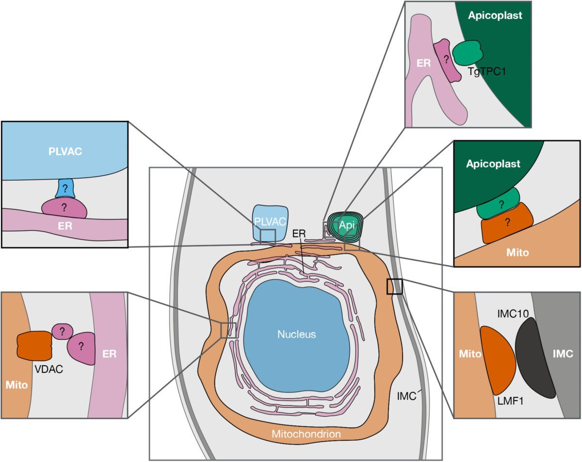
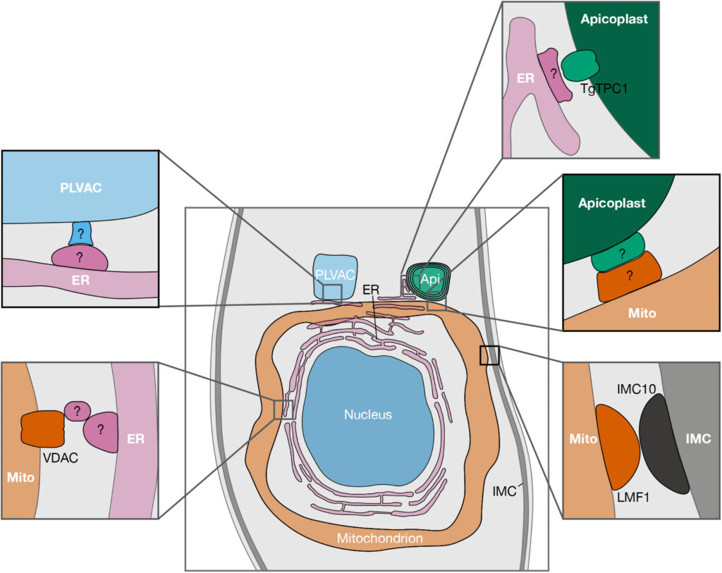
Figure 1. Reported and potential MCSs between organelles of Toxoplasma gondii. Schematic representation showing proteins recently reported to be involved in MCSs, along with putative MCS candidates (indicated with “?”). For clarity purposes, only the central part of the parasite is shown. Abbreviations: AP, apicoplast; ER, endoplasmic reticulum; PLVAC, plant-like vacuolar compartment; IMC, inner membrane complex; TgTPC, T. gondii two pore channel; VDAC, voltage-dependent anion channel; LMF1, lasso maintenance factor 1.; MCS, membrane contact site.


Apicomplexan parasites are a group of protists that cause disease in humans and include pathogens like Plasmodium spp., the causative agent of malaria, and Toxoplasma gondii, the etiological agent of toxoplasmosis and one of the most ubiquitous human parasites in the world. Membrane contact sites (MCSs) are widespread structures within eukaryotic cells but their characterization in apicomplexan parasites is only in its very beginnings. Basic biological features of the T. gondii parasitic cycle support numerous organellar interactions, including the transfer of Ca2+ and metabolites between different compartments. In T. gondii, Ca2+ signals precede a series of interrelated molecular processes occurring in a coordinated manner that culminate in the stimulation of key steps of the parasite life cycle. Calcium transfer from the endoplasmic reticulum to other organelles via MCSs would explain the precision, speed, and efficiency that is needed during the lytic cycle of T. gondii. In this short review, we discuss the implications of these structures in cellular signaling, with an emphasis on their potential role in Ca2+ signaling.

Diego Huet, Silvia N J Moreno. Contact (Thousand Oaks). 2023 Aug 6;6:25152564231189064. doi: 10.1177/25152564231189064. eCollection 2023 Jan-Dec.

Analysis of the Interactome of the Toxoplasma gondii Tgj1 HSP40 Chaperone

Toxoplasma gondii is an obligate intracellular apicomplexan that causes toxoplasmosis in humans and animals. Central to its dissemination and pathogenicity is the ability to rapidly divide in the tachyzoite stage and infect any type of nucleated cell. Adaptation to different cell contexts requires high plasticity in which heat shock proteins (Hsps) could play a fundamental role. Tgj1 is a type I Hsp40 of T. gondii, an ortholog of the DNAJA1 group, which is essential during the tachyzoite lytic cycle. Tgj1 consists of a J-domain, ZFD, and DNAJ_C domains with a CRQQ C-terminal motif, which is usually prone to lipidation. Tgj1 presented a mostly cytosolic subcellular localization overlapping partially with endoplasmic reticulum. Protein-protein Interaction (PPI) analysis showed that Tgj1 could be implicated in various biological pathways, mainly translation, protein folding, energy metabolism, membrane transport and protein translocation, invasion/pathogenesis, cell signaling, chromatin and transcription regulation, and cell redox homeostasis among others. The combination of Tgj1 and Hsp90 PPIs retrieved only 70 interactors linked to the Tgj1-Hsp90 axis, suggesting that Tgj1 would present specific functions in addition to those of the Hsp70/Hsp90 cycle, standing out invasion/pathogenesis, cell shape motility, and energy pathway. Within the Hsp70/Hsp90 cycle, translation-associated pathways, cell redox homeostasis, and protein folding were highly enriched in the Tgj1-Hsp90 axis. In conclusion, Tgj1 would interact with a wide range of proteins from different biological pathways, which could suggest a relevant role in them.

Jonathan Munera López, Andrés Mariano Alonso, Maria Julia Figueras, Ana María Saldarriaga Cartagena, Miryam A Hortua Triana, Luis Diambra, Laura Vanagas, Bin Deng, Silvia N J Moreno, Sergio Oscar Angel. Proteomes. 2023 Mar 1;11(1):9. doi: 10.3390/proteomes11010009.

The Toxoplasma Plant-Like Vacuolar Compartment (PLVAC)

Toxoplasma gondii belongs to the phylum Apicomplexa and is an important cause of congenital disease and infection in immunocompromised patients. T. gondii shares several characteristics with plants including a non-photosynthetic plastid termed apicoplast and a multi-vesicular organelle that was named the plant-like vacuole (PLV) or vacuolar compartment (VAC). The name plant-like vacuole was selected based on its resemblance in composition and function to plant vacuoles. The name VAC represents its general vacuolar characteristics. We will refer to the organelle as PLVAC in this review. New findings in recent years have revealed that the PLVAC represents the lysosomal compartment of T. gondii which has adapted peculiarities to fulfill specific Toxoplasma needs. In this review, we discuss the composition and functions of the PLVAC highlighting its roles in ion storage and homeostasis, endocytosis, exocytosis, and autophagy.

Andrew J Stasic, Silvia N J Moreno, Vern B Carruthers, Zhicheng Dou. J Eukaryot Microbiol. 2022 Oct 11;e12951. doi: 10.1111/jeu.12951.

The Heptaprenyl Diphosphate Synthase (Coq1) Is the Target of a Lipophilic Bisphosphonate That Protects Mice against Toxoplasma gondii Infection

Prenyldiphosphate synthases catalyze the reaction of allylic diphosphates with one or more isopentenyl diphosphate molecules to form compounds such as farnesyl diphosphate, used in, e.g., sterol biosynthesis and protein prenylation, as well as longer “polyprenyl” diphosphates, used in ubiquinone and menaquinone biosynthesis. Quinones play an essential role in electron transport and are associated with the inner mitochondrial membrane due to the presence of the polyprenyl group. In this work, we investigated the synthesis of the polyprenyl diphosphate that alkylates the ubiquinone ring precursor in Toxoplasma gondii, an opportunistic pathogen that causes serious disease in immunocompromised patients and the unborn fetus. The enzyme that catalyzes this early step of the ubiquinone synthesis is Coq1 (TgCoq1), and we show that it produces the C35 species heptaprenyl diphosphate. TgCoq1 localizes to the mitochondrion and is essential for in vitro T. gondii growth. We demonstrate that the growth defect of a T. gondii TgCoq1 mutant is rescued by complementation with a homologous TgCoq1 gene or with a (C45) solanesyl diphosphate synthase from Trypanosoma cruzi (TcSPPS). We find that a lipophilic bisphosphonate (BPH-1218) inhibits T. gondii growth at low-nanomolar concentrations, while overexpression of the TgCoq1 enzyme dramatically reduced growth inhibition by the bisphosphonate. Both the severe growth defect of the mutant and the inhibition by BPH-1218 were rescued by supplementation with a long-chain (C30) ubiquinone (UQ6). Importantly, BPH-1218 also protected mice against a lethal T. gondii infection. TgCoq1 thus represents a potential drug target that could be exploited for improved chemotherapy of toxoplasmosis.

IMPORTANCE Millions of people are infected with Toxoplasma gondii, and the available treatment for toxoplasmosis is not ideal. Most of the drugs currently used are only effective for the acute infection, and treatment can trigger serious side effects requiring changes in the therapeutic approach. There is, therefore, a compelling need for safe and effective treatments for toxoplasmosis. In this work, we characterize an enzyme of the mitochondrion of T. gondii that can be inhibited by an isoprenoid pathway inhibitor. We present evidence that demonstrates that inhibition of the enzyme is linked to parasite death. In addition, the inhibitor can protect mice against a lethal dose of T. gondii. Our results thus reveal a promising chemotherapeutic target for the development of new medicines for toxoplasmosis.

Melissa A Sleda, Zhu-Hong Li, Ranjan Behera, Baihetiya Baierna, Catherine Li, Jomkwan Jumpathong, Satish R Malwal, Makoto Kawamukai, Eric Oldfield, Silvia N J Moreno. mBio. 2022 Sep 21;e0196622. doi: 10.1128/mbio.01966-22.

Disruption of Toxoplasma gondii-Induced Host Cell DNA Replication Is Dependent on Contact Inhibition and Host Cell Type

The protozoan Toxoplasma gondii is a highly successful obligate intracellular parasite that, upon invasion of its host cell, releases an array of host-modulating protein effectors to counter host defenses and further its own replication and dissemination. Early studies investigating the impact of T. gondii infection on host cell function revealed that this parasite can force normally quiescent cells to activate their cell cycle program. Prior reports by two independent groups identified the dense granule protein effector HCE1/TEEGR as being solely responsible for driving host cell transcriptional changes through its direct interaction with the cyclin E regulatory complex DP1 and associated transcription factors. Our group independently identified HCE1/TEEGR through the presence of distinct repeated regions found in a number of host nuclear targeted parasite effectors and verified its central role in initiating host cell cycle changes. Additionally, we report here the time-resolved kinetics of host cell cycle transition in response to HCE1/TEEGR, using the fluorescence ubiquitination cell cycle indicator reporter line (FUCCI), and reveal the existence of a block in S-phase progression and host DNA synthesis in several cell lines commonly used in the study of T. gondii. Importantly, we have observed that this S-phase block is not due to additional dense granule effectors but rather is dependent on the host cell line background and contact inhibition status of the host monolayer in vitro. This work highlights intriguing differences in the host response to reprogramming by the parasite and raises interesting questions regarding how parasite effectors differentially manipulate the host cell depending on the in vitro or in vivo context.

IMPORTANCE Toxoplasma gondii chronically infects approximately one-third of the global population and can produce severe pathology in immunologically immature or compromised individuals. During infection, this parasite releases numerous host-targeted effector proteins that can dramatically alter the expression of a variety of host genes. A better understanding of parasite effectors and their host targets has the potential to not only provide ways to control infection but also inform us about our own basic biology. One host pathway that has been known to be altered by T. gondii infection is the cell cycle, and prior reports have identified a parasite effector, known as HCE1/TEEGR, as being responsible. In this report, we further our understanding of the kinetics of cell cycle transition induced by this effector and show that the capacity of HCE1/TEEGR to induce host cell DNA synthesis is dependent on both the cell type and the status of contact inhibition.

Edwin Pierre-Louis, Menna G Etheridge, Rodrigo de Paula Baptista, Asis Khan, Nathan M Chasen, Ronald D Etheridge. mSphere. 2022 May 19;e0016022. doi: 10.1128/msphere.00160-22.

Parasite Powerhouse: a Review of the Toxoplasma gondii Mitochondrion

Toxoplasma gondii is a member of the apicomplexan phylum, a group of single-celled eukaryotic parasites that cause significant human morbidity and mortality around the world. T. gondii harbors two organelles of endosymbiotic origin: a non-photosynthetic plastid, known as the apicoplast, and a single mitochondrion derived from the ancient engulfment of an α-proteobacterium. Due to excitement surrounding the novelty of the apicoplast, the T. gondii mitochondrion was, to a certain extent, overlooked for about two decades. However, recent work has illustrated that the mitochondrion is an essential hub of apicomplexan-specific biology. Development of novel techniques, such as cryo-electron microscopy, complexome profiling, and next-generation sequencing have led to a renaissance in mitochondrial studies. This review will cover what is currently known about key features of the T. gondii mitochondrion, ranging from its genome to protein import machinery and biochemical pathways. Particular focus will be given to mitochondrial features that diverge significantly from the mammalian host, along with discussion of this important organelle as a drug target.

Madelaine M. Usey, Diego Huet. J Eukaryot Microbiol. 2022 Mar 21;e12906. doi: 10.1111/jeu.12906.

In Vivo Efficacy of SQ109 against Leishmania donovani, Trypanosoma spp. and Toxoplasma gondii and In Vitro Activity of SQ109 Metabolites

SQ109 is an anti-tubercular drug candidate that has completed Phase IIb/III clinical trials for tuberculosis and has also been shown to exhibit potent in vitro efficacy against protozoan parasites including Leishmania and Trypanosoma cruzi spp. However, its in vivo efficacy against protozoa has not been reported. Here, we evaluated the activity of SQ109 in mouse models of Leishmania, Trypanosoma spp. as well as Toxoplasma infection. In the T. cruzi mouse model, 80% of SQ109-treated mice survived at 40 days post-infection. Even though SQ109 did not cure all mice, these results are of interest since they provide a basis for future testing of combination therapies with the azole posaconazole, which acts synergistically with SQ109 in vitro. We also found that SQ109 inhibited the growth of Toxoplasma gondii in vitro with an IC50 of 1.82 µM and there was an 80% survival in mice treated with SQ109, whereas all untreated animals died 10 days post-infection. Results with Trypanosoma brucei and Leishmania donovani infected mice were not promising with only moderate efficacy. Since SQ109 is known to be extensively metabolized in animals, we investigated the activity in vitro of SQ109 metabolites. Among 16 metabolites, six mono-oxygenated forms were found active across the tested protozoan parasites, and there was a ~6× average decrease in activity of the metabolites as compared to SQ109 which is smaller than the ~25× found with mycobacteria.

Kyung-Hwa Baek, Trong-Nhat Phan, Satish R Malwal, Hyeryon Lee, Zhu-Hong Li, Silvia N J Moreno, Eric Oldfield, Joo Hwan No. Biomedicines. 2022 Mar 14;10(3):670. doi: 10.3390/biomedicines10030670.

A plastid two-pore channel essential for inter-organelle communication and growth of Toxoplasma gondii

Two-pore channels (TPCs) are a ubiquitous family of cation channels that localize to acidic organelles in animals and plants to regulate numerous Ca2+-dependent events. Little is known about TPCs in unicellular organisms despite their ancient origins. Here, we characterize a TPC from Toxoplasma gondii, the causative agent of toxoplasmosis. TgTPC is a member of a novel clad of TPCs in Apicomplexa, distinct from previously identified TPCs and only present in coccidians. We show that TgTPC localizes not to acidic organelles but to the apicoplast, a non-photosynthetic plastid found in most apicomplexan parasites. Conditional silencing of TgTPC resulted in progressive loss of apicoplast integrity, severely affecting growth and the lytic cycle. Isolation of TPC null mutants revealed a selective role for TPCs in replication independent of apicoplast loss that required conserved residues within the pore-lining region. Using a genetically-encoded Ca2+ indicator targeted to the apicoplast, we show that Ca2+ signals deriving from the ER but not from the extracellular space are selectively transmitted to the lumen. Deletion of the TgTPC gene caused reduced apicoplast Ca2+ uptake and membrane contact site formation between the apicoplast and the ER. Fundamental roles for TPCs in maintaining organelle integrity, inter-organelle communication and growth emerge.

Zhu-Hong Li, Thayer P King, Lawrence Ayong, Beejan Asady, Xinjiang Cai, Taufiq Rahman, Stephen A Vella, Isabelle Coppens, Sandip Patel, Silvia N J Moreno. Nat Commun. 2021 Oct 4;12(1):5802. doi: 10.1038/s41467-021-25987-5

Calcium signaling through a Transient Receptor Channel is important for Toxoplasma gondii growth

Transient Receptor Potential (TRP) channels participate in calcium ion (Ca2+) influx and intracellular Ca2+ release. TRP channels have not been studied in Toxoplasma gondii or any other apicomplexan parasite. In this work we characterize TgGT1_310560, a protein predicted to possess a TRP domain (TgTRPPL-2) and determined its role in Ca2+ signaling in T. gondii, the causative agent of toxoplasmosis. TgTRPPL-2 localizes to the plasma membrane and the endoplasmic reticulum (ER) of T. gondii. The ΔTgTRPPL-2 mutant was defective in growth and cytosolic Ca2+ influx from both extracellular and intracellular sources. Heterologous expression of TgTRPPL-2 in HEK-3KO cells allowed its functional characterization. Patching of ER-nuclear membranes demonstrates that TgTRPPL-2 is a non-selective cation channel that conducts Ca2+. Pharmacological blockers of TgTRPPL-2 inhibit Ca2+ influx and parasite growth. This is the first report of an apicomplexan ion channel that conducts Ca2+ and may initiate a Ca2+ signaling cascade that leads to the stimulation of motility, invasion and egress. TgTRPPL-2 is a potential target for combating Toxoplasmosis.

Karla Marie Marquez-Nogueras, Myriam Andrea Hortua Triana, Nathan M Chasen, Ivana Y Kuo, Silvia NJ Moreno. Elife. 2021 Jun 9;10:e63417. doi: 10.7554/eLife.63417.

Ca2+ entry at the plasma membrane and uptake by acidic stores is regulated by the activity of the V‐H+‐ATPase in Toxoplasma gondii

Ca2+ is a universal intracellular signal that regulates many cellular functions. In Toxoplasma gondii, the controlled influx of extracellular and intracellular Ca2+ into the cytosol initiates a signaling cascade that promotes pathogenic processes like tissue destruction and dissemination. In this work we studied the role of proton transport in cytosolic Ca2+ homeostasis and the initiation of Ca2+ signaling. We used a T. gondii mutant of the V-ATPase, a pump previously shown to transport protons to the extracellular medium, control intracellular pH and membrane potential and we show that proton gradients are important for maintaining resting cytosolic Ca2+ at physiological levels and for Ca2+ influx. Proton transport was also important for Ca2+ storage by acidic stores and, unexpectedly, the endoplasmic reticulum. Proton transport impacted the amount of polyphosphate (polyP), a phosphate polymer that binds Ca2+ and concentrate in acidocalcisomes. This was supported by the co-localization of the vacuolar transporter chaperone 4 (VTC4), the catalytic subunit of the VTC complex that synthesizes polyP, with the V-ATPase in acidocalcisomes. Our work show that proton transport regulate plasma membrane Ca2+ transport and control acidocalcisome polyP and Ca2+ content impacting Ca2+ signaling and downstream stimulation of motility and egress in T. gondii.

Andrew J Stasic, Eric J Dykes, Ciro D Cordeiro, Stephen A Vella, Mojtaba S Fazli, Shannon Quinn, Roberto Docampo, Silvia N J Moreno. Mol Microbiol. 2021 Apr 1. doi: 10.1111/mmi.14722