Generic selectors
Exact matches only
Search in title
Search in content
Post Type Selectors

Tag: Toxoplasma gondii

Two CTEGD trainees receive AHA fellowships
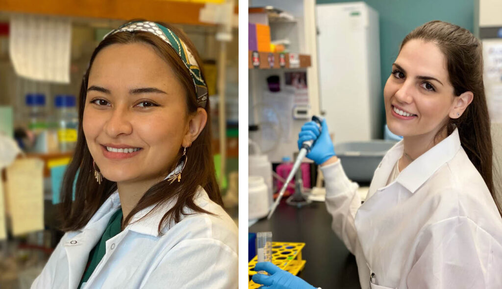
Photos of Graduate student Baihetiya “Barna” Baierna and postdoctoral fellow Mayara Bertolini
Graduate student Baihetiya “Barna” Baierna and postdoctoral fellow Mayara Bertolini received fellowships from the American Heart Association, supporting their research and education. Both are studying parasites in the University of Georgia’s Center for Tropical and Emerging Global Diseases. (Photos courtesy of CTEGD)


Baihetiya “Barna” Baierna, a cellular biology graduate student in Silvia Moreno’s laboratory, received an American Heart Association Pre-doctoral Fellowship. It will fund her training for the next two years as she studies the mitochondrion of Toxoplasma gondii.

Baierna grew up wanting to follow in her mother’s footsteps as a scientist.

“My mom worked for the regional CDC in China and I was interested in science since a young age,” Baierna said.

After completing her undergraduate degree in biochemistry, she was sure she wanted to continue her training in graduate school. After being accepted into the Department of Cellular Biology program, she joined the Moreno Laboratory.

Toxoplasma gondii infects approximately one third of the world human population. The infection can cause serious complications in people with a suppressed immune system. Baierna’s research aims at validating novel T. gondii mitochondrial proteins as novel chemotherapeutic targets for improved chemotherapy of toxoplasmosis. This is important because the present drugs are not effective against the chronic stages of the infection. She has developed novel strategies for the discovery of new mitochondrial proteins and already found a novel enzymatic activity highly divergent from the mammalian counterpart. The outcome of this project will expand the knowledge of the T. gondii mitochondrion, as well as helping with the identification of viable drug targets.

“An AHA Fellowship is a very competitive award, but Barna deserves it and we are very proud of her,” said Moreno.

“Preparing the grant proposal was a great learning experience and it will help me with my career development,” said Baierna, “I’m very happy that it was funded.”

Mayara Bertolini, a post-doctoral fellow in Roberto Docampo’s laboratory, received an American Heart Association Post-doctoral Fellowship. It will support her training for one year.

After receiving her bachelor’s degree, Bertolini obtained her master’s degree in a lab that Docampo had set up in Brazil working on T. cruzi. From there she decided to pursue her Ph.D. at the University of Georgia. She completed her Ph.D. in 2023.

Trypanosoma cruzi is the parasite that causes Chagas disease. At least 6 million people, mostly in South America, are infected with the parasite. T. cruzi is transmitted to humans through the feces of an insect commonly referred to as the kissing bug. While Chagas disease was first discovered in 1909, there is still a lot that is unknown about the biology of T. cruzi. This lack of knowledge has hindered drug development. Bertolini’s project is focused on the role of polyphosphate during the Trypanosoma cruzi life cycle.

“This is the second fellowship from the AHA that Mayara has received. She got a two-year pre-doctoral fellowship before and has done outstanding work,” said Docampo.

“AHA Fellowships are very competitive and I’m thrilled my proposal was selected,” said Bertolini. “In addition to supporting my training, there is support for career development and networking opportunities.”


The story originally appeared at https://research.uga.edu/news/two-ctegd-trainees-receive-aha-fellowships/

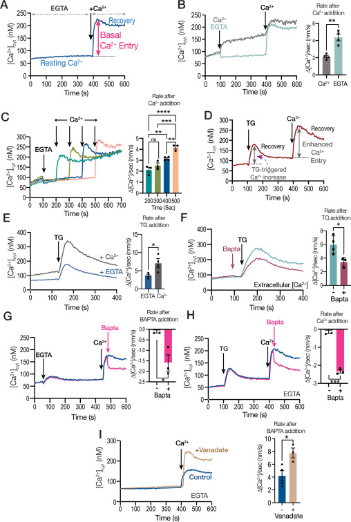
Regulation of Calcium entry by cyclic GMP signaling in Toxoplasma gondii

Figure 1. Calcium entry through the plasma membrane of extracellular T. gondii tachyzoites.


Ca2+ signaling impacts almost every aspect of cellular life. Ca2+ signals are generated through the opening of ion channels that permit the flow of Ca2+ down an electrochemical gradient. Cytosolic Ca2+ fluctuations can be generated through Ca2+ entry from the extracellular milieu or release from intracellular stores. In Toxoplasma gondii, Ca2+ ions play critical roles in several essential functions for the parasite like invasion of host cells, motility and egress. Plasma membrane Ca2+ entry in T. gondii was previously shown to be activated by cytosolic calcium and inhibited by the voltage-operated Ca2+ channel blocker nifedipine. However, Ca2+ entry in T. gondii did not show the classical characteristics of store regulation. In this work, we characterized the mechanism by which cytosolic Ca2+ regulates plasma membrane Ca2+ entry in extracellular T. gondii tachyzoites loaded with the Ca2+ indicator Fura 2. We compared the inhibition by nifedipine with the effect of the broad spectrum TRP channel inhibitor, anthranilic acid or ACA and we find that both inhibitors act on different Ca2+ entry activities. We demonstrate, using pharmacological and genetic tools, that an intracellular signaling pathway engaging cyclicGMP (cGMP), protein kinase G (PKG), Ca2+ and the phosphatidyl inositol phospholipase C (PI-PLC) affects Ca2+ entry and we present a model for crosstalk between cGMP and cytosolic Ca2+ for the activation of T. gondii‘s lytic cycle traits.

Miryam A Hortua Triana, Karla M Márquez-Nogueras, Mojtaba Sedigh Fazli, Shannon Quinn, Silvia N J Moreno. J Biol Chem. 2024 Feb 19:105771. doi: 10.1016/j.jbc.2024.105771

Diego Huet zeroes in on parasite that affects thousands each year

Diego Huet
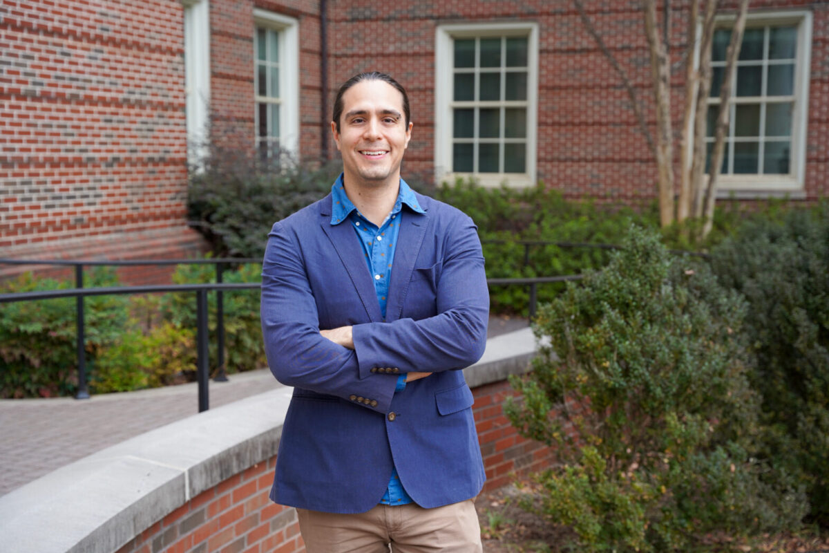
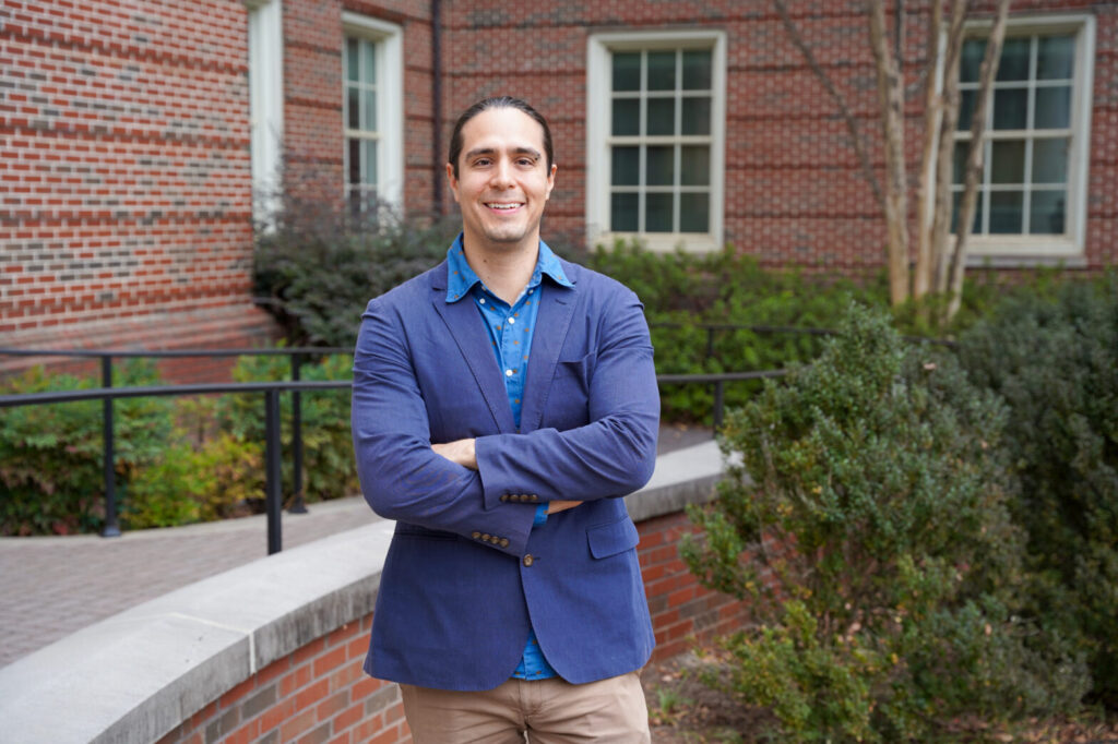
Diego Huet, assistant professor in the College of Pharmacy and the Center for Tropical & Emerging Global Diseases, studies parasites that cause disease in both humans and animals. His lab has ramped up a project to better understand the biology of Toxoplasma gondii , an organism carried by cats that is related to the parasite that causes malaria. (Photo by Lauren Corcino)


From an early age, Diego Huet has been interested in the unusual and fascinating found in the natural world.

His early encounters with animals, plants and insects nurtured his curiosity about nature. Their striking colors and sometimes strange shapes drew his interest, and even today he continues to capture them through macro photography. It was this fascination that led him to the parasite he studies today.

“I was always drawn to ‘unconventional’ or ‘weird’ science,” said Huet, an assistant professor in the College of Pharmacy’s Department of Pharmaceutical and Biomedical Sciences and member of the Center for Tropical and Emerging Global Diseases. “But I also wanted to be hands on, which is what led me to molecular and cellular biology.”

As a doctoral student, Huet began studying Trypanosoma brucei, a parasite commonly transmitted by the tsetse fly. Wanting to study a different parasite as a postdoctoral researcher, he was torn between studying Plasmodium, which is the causative agent of malaria, and Toxoplasma gondii, a related parasite that is carried by cats. Both parasites, which belong to a group of organisms called apicomplexans, cause diseases in humans and animals, and there remain large knowledge gaps in our basic understanding of them. Ultimately, he chose the latter.

Plasmodium is difficult to manipulate,” Huet said. “Toxoplasma is related to Plasmodium, but is easier to work with because it isn’t as complex, and what we learn about Toxo could also increase our knowledge of Plasmodium.”

Just as yeast and fruit flies are used as model organisms to study human biology, Toxoplasma can be used as a model for shared features of apicomplexan biology.

Besides aiding in the understanding of other parasites of human and veterinary concern, including parasites that cause malaria in tropical and subtropical regions of the world, Toxoplasma gondii also causes human and animal disease. More than 40 million people in the U.S. are estimated to carry T. gondii. Although most never show symptoms, it poses a major health threat to immunocompromised individuals and pregnant women as it can lead to miscarriage and birth defects. Toxoplasmosis, the disease caused by Toxoplasma, is considered a leading cause of death among foodborne illnesses though it can also be transmitted through contact with cat feces.

The Centers for Disease Control and Prevention has it listed as a neglected parasitic infection in the United States and a target for public health action.

Huet joined the faculty at the University of Georgia in 2019 and has developed a robust research program to expand knowledge of the basic biology of Toxoplasma.

Madelaine Usey
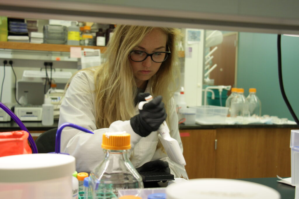
Madelaine Usey is a cellular biology graduate student in the Huet Laboratory.

In a recently published study in “mBio”, cellular biology doctoral candidate and Huet Lab member Madelaine Usey looked at proteins critical for mitochondrial function in T. gondii. The mitochondrion is considered the “powerhouse of the cell,” but it is an enzyme called ATP synthase that generates the cellular energy.

“Our findings are really exciting for drug discovery,” Usey said. “Many of the proteins that make up the ATP synthase are different in Toxoplasma compared to other organisms. In this study, we were able to figure out what two of those novel subunits are doing—they act as scaffolding for this enormous ATP synthase complex.”

These proteins are unique to Toxoplasma and could be used in drug discovery as targets since they are important for mitochondrial functioning.

Another project in Huet’s laboratory, which recently received funding through a grant from the National Institute of General Medical Sciences, investigates how organelles within the parasite communicate.

“Traditionally, we thought organelles send and receive calcium and other metabolites in much the same way we receive a package through the mail,” Huet said. “Cells form vesicles to transport materials to specific locations within the cell. The vesicles are labeled with proteins that act like a postal address, telling the vesicle where to go.”

However, cells can also exchange material through another process.

“When the organelles’ membranes get close together, they form what is called a membrane contact site,” Huet said. “In this case it is more like one organelle hand delivers the package to another.”

A membrane contact site is a specialized protein structure that organelles use for intracellular communication. However, it is not a well understood structure in apicomplexans. In addition, these parasites have additional organelles not found in traditional models like humans and yeast, so Huet is trying to understand how the organellar communication is happening in apicomplexans using Toxoplasma as a model.

Identifying such proteins and their functions could lead to better drug targets and better drug treatments, which all the neglected parasitic diseases need.

“Toxo’s genome isn’t well annotated,” Huet said. “Finding membrane contact site proteins is an arduous task—it’s a goal of my lab to identify some of them and their involvement in Toxoplasma membrane contact sites.”


This article was first published at https://research.uga.edu/news/diego-huet-zeroes-in-on-parasite-that-affects-thousands-each-year/

ATP synthase-associated coiled-coil-helix-coiled-coil-helix (CHCH) domain-containing proteins are critical for mitochondrial function in Toxoplasma gondii

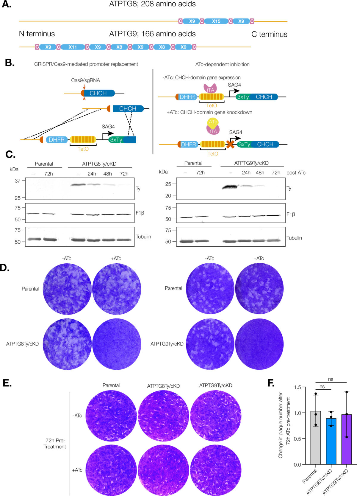
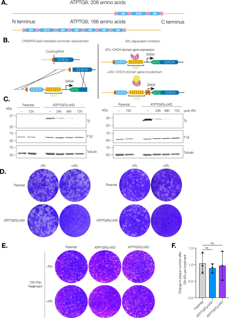
CHCH domain proteins associated with the T. gondii ATP synthase are essential for the lytic cycle. (A) Schematic representation of the CHCH domain size and location in ATPTG8 and ATPTG9. C represents cysteine residues and X represents any other amino acid residue.

Coiled-coil-helix-coiled-coil-helix (CHCH) domains consist of two pairs of cysteine residues that are oxidized to form disulfide bonds upon mitochondrial import. Proteins containing these domains play important roles in mitochondrial ultrastructure and in the biogenesis, function, and stability of electron transport chain complexes. Interestingly, recent investigations of the Toxoplasma gondii ATP synthase identified subunits containing CHCH domains. As CHCH domain proteins have never been found in any other ATP synthase, their role in T. gondii was unclear. Using conditional gene knockdown systems, we investigated two T. gondii ATP synthase subunits containing CHCH domains: ATPTG8 and ATPTG9. We show that these two subunits are essential for the lytic cycle as well as stability and function of the ATP synthase. Further, we illustrated that their knockdown disrupts multiple aspects of mitochondrial morphology, including ultrastructure and cristae density. Mutation of key cysteine residues in the CHCH domains also caused mis-localization of the proteins. Our work suggests that these proteins likely provide structural support to the exceptionally large T. gondii ATP synthase complex and that perturbations to the structural integrity of this complex result in deleterious downstream effects on the parasite mitochondrion. These investigations add to a growing body of work focused on the divergent aspects of the apicomplexan ATP synthase, which could ultimately uncover novel drug targets. IMPORTANCE Members of the coiled-coil-helix-coiled-coil-helix (CHCH) domain protein family are transported into the mitochondrial intermembrane space, where they play important roles in the biogenesis and function of the organelle. Unexpectedly, the ATP synthase of the apicomplexan Toxoplasma gondii harbors CHCH domain-containing subunits of unknown function. As no other ATP synthase studied to date contains this class of proteins, characterizing their function will be of broad interest to the fields of molecular parasitology and mitochondrial evolution. Here, we demonstrate that that two T. gondii ATP synthase subunits containing CHCH domains are required for parasite survival and for stability and function of the ATP synthase. We also show that knockdown disrupts multiple aspects of the mitochondrial morphology of T. gondii and that mutation of key residues in the CHCH domains caused mis-localization of the proteins. This work provides insight into the unique features of the apicomplexan ATP synthase, which could help to develop therapeutic interventions against this parasite and other apicomplexans, such as the malaria-causing parasite Plasmodium falciparum.

Madelaine M Usey, Diego Huet. mBio. 2023 Oct 5:e0176923. doi: 10.1128/mbio.01769-23.

Interorganellar Communication Through Membrane Contact Sites in Toxoplasma gondii

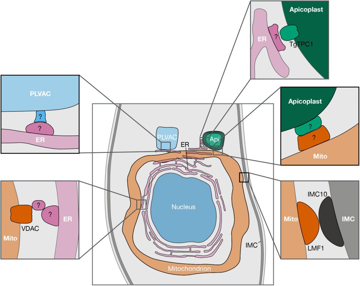
Figure 1. Reported and potential MCSs between organelles of Toxoplasma gondii. Schematic representation showing proteins recently reported to be involved in MCSs, along with putative MCS candidates (indicated with “?”). For clarity purposes, only the central part of the parasite is shown. Abbreviations: AP, apicoplast; ER, endoplasmic reticulum; PLVAC, plant-like vacuolar compartment; IMC, inner membrane complex; TgTPC, T. gondii two pore channel; VDAC, voltage-dependent anion channel; LMF1, lasso maintenance factor 1.; MCS, membrane contact site.


Apicomplexan parasites are a group of protists that cause disease in humans and include pathogens like Plasmodium spp., the causative agent of malaria, and Toxoplasma gondii, the etiological agent of toxoplasmosis and one of the most ubiquitous human parasites in the world. Membrane contact sites (MCSs) are widespread structures within eukaryotic cells but their characterization in apicomplexan parasites is only in its very beginnings. Basic biological features of the T. gondii parasitic cycle support numerous organellar interactions, including the transfer of Ca2+ and metabolites between different compartments. In T. gondii, Ca2+ signals precede a series of interrelated molecular processes occurring in a coordinated manner that culminate in the stimulation of key steps of the parasite life cycle. Calcium transfer from the endoplasmic reticulum to other organelles via MCSs would explain the precision, speed, and efficiency that is needed during the lytic cycle of T. gondii. In this short review, we discuss the implications of these structures in cellular signaling, with an emphasis on their potential role in Ca2+ signaling.

Diego Huet, Silvia N J Moreno. Contact (Thousand Oaks). 2023 Aug 6;6:25152564231189064. doi: 10.1177/25152564231189064. eCollection 2023 Jan-Dec.

Analysis of the Interactome of the Toxoplasma gondii Tgj1 HSP40 Chaperone

Toxoplasma gondii is an obligate intracellular apicomplexan that causes toxoplasmosis in humans and animals. Central to its dissemination and pathogenicity is the ability to rapidly divide in the tachyzoite stage and infect any type of nucleated cell. Adaptation to different cell contexts requires high plasticity in which heat shock proteins (Hsps) could play a fundamental role. Tgj1 is a type I Hsp40 of T. gondii, an ortholog of the DNAJA1 group, which is essential during the tachyzoite lytic cycle. Tgj1 consists of a J-domain, ZFD, and DNAJ_C domains with a CRQQ C-terminal motif, which is usually prone to lipidation. Tgj1 presented a mostly cytosolic subcellular localization overlapping partially with endoplasmic reticulum. Protein-protein Interaction (PPI) analysis showed that Tgj1 could be implicated in various biological pathways, mainly translation, protein folding, energy metabolism, membrane transport and protein translocation, invasion/pathogenesis, cell signaling, chromatin and transcription regulation, and cell redox homeostasis among others. The combination of Tgj1 and Hsp90 PPIs retrieved only 70 interactors linked to the Tgj1-Hsp90 axis, suggesting that Tgj1 would present specific functions in addition to those of the Hsp70/Hsp90 cycle, standing out invasion/pathogenesis, cell shape motility, and energy pathway. Within the Hsp70/Hsp90 cycle, translation-associated pathways, cell redox homeostasis, and protein folding were highly enriched in the Tgj1-Hsp90 axis. In conclusion, Tgj1 would interact with a wide range of proteins from different biological pathways, which could suggest a relevant role in them.

Jonathan Munera López, Andrés Mariano Alonso, Maria Julia Figueras, Ana María Saldarriaga Cartagena, Miryam A Hortua Triana, Luis Diambra, Laura Vanagas, Bin Deng, Silvia N J Moreno, Sergio Oscar Angel. Proteomes. 2023 Mar 1;11(1):9. doi: 10.3390/proteomes11010009.

The Toxoplasma Plant-Like Vacuolar Compartment (PLVAC)

Toxoplasma gondii belongs to the phylum Apicomplexa and is an important cause of congenital disease and infection in immunocompromised patients. T. gondii shares several characteristics with plants including a non-photosynthetic plastid termed apicoplast and a multi-vesicular organelle that was named the plant-like vacuole (PLV) or vacuolar compartment (VAC). The name plant-like vacuole was selected based on its resemblance in composition and function to plant vacuoles. The name VAC represents its general vacuolar characteristics. We will refer to the organelle as PLVAC in this review. New findings in recent years have revealed that the PLVAC represents the lysosomal compartment of T. gondii which has adapted peculiarities to fulfill specific Toxoplasma needs. In this review, we discuss the composition and functions of the PLVAC highlighting its roles in ion storage and homeostasis, endocytosis, exocytosis, and autophagy.

Andrew J Stasic, Silvia N J Moreno, Vern B Carruthers, Zhicheng Dou. J Eukaryot Microbiol. 2022 Oct 11;e12951. doi: 10.1111/jeu.12951.

The Heptaprenyl Diphosphate Synthase (Coq1) Is the Target of a Lipophilic Bisphosphonate That Protects Mice against Toxoplasma gondii Infection

Prenyldiphosphate synthases catalyze the reaction of allylic diphosphates with one or more isopentenyl diphosphate molecules to form compounds such as farnesyl diphosphate, used in, e.g., sterol biosynthesis and protein prenylation, as well as longer “polyprenyl” diphosphates, used in ubiquinone and menaquinone biosynthesis. Quinones play an essential role in electron transport and are associated with the inner mitochondrial membrane due to the presence of the polyprenyl group. In this work, we investigated the synthesis of the polyprenyl diphosphate that alkylates the ubiquinone ring precursor in Toxoplasma gondii, an opportunistic pathogen that causes serious disease in immunocompromised patients and the unborn fetus. The enzyme that catalyzes this early step of the ubiquinone synthesis is Coq1 (TgCoq1), and we show that it produces the C35 species heptaprenyl diphosphate. TgCoq1 localizes to the mitochondrion and is essential for in vitro T. gondii growth. We demonstrate that the growth defect of a T. gondii TgCoq1 mutant is rescued by complementation with a homologous TgCoq1 gene or with a (C45) solanesyl diphosphate synthase from Trypanosoma cruzi (TcSPPS). We find that a lipophilic bisphosphonate (BPH-1218) inhibits T. gondii growth at low-nanomolar concentrations, while overexpression of the TgCoq1 enzyme dramatically reduced growth inhibition by the bisphosphonate. Both the severe growth defect of the mutant and the inhibition by BPH-1218 were rescued by supplementation with a long-chain (C30) ubiquinone (UQ6). Importantly, BPH-1218 also protected mice against a lethal T. gondii infection. TgCoq1 thus represents a potential drug target that could be exploited for improved chemotherapy of toxoplasmosis.

IMPORTANCE Millions of people are infected with Toxoplasma gondii, and the available treatment for toxoplasmosis is not ideal. Most of the drugs currently used are only effective for the acute infection, and treatment can trigger serious side effects requiring changes in the therapeutic approach. There is, therefore, a compelling need for safe and effective treatments for toxoplasmosis. In this work, we characterize an enzyme of the mitochondrion of T. gondii that can be inhibited by an isoprenoid pathway inhibitor. We present evidence that demonstrates that inhibition of the enzyme is linked to parasite death. In addition, the inhibitor can protect mice against a lethal dose of T. gondii. Our results thus reveal a promising chemotherapeutic target for the development of new medicines for toxoplasmosis.

Melissa A Sleda, Zhu-Hong Li, Ranjan Behera, Baihetiya Baierna, Catherine Li, Jomkwan Jumpathong, Satish R Malwal, Makoto Kawamukai, Eric Oldfield, Silvia N J Moreno. mBio. 2022 Sep 21;e0196622. doi: 10.1128/mbio.01966-22.

Disruption of Toxoplasma gondii-Induced Host Cell DNA Replication Is Dependent on Contact Inhibition and Host Cell Type

The protozoan Toxoplasma gondii is a highly successful obligate intracellular parasite that, upon invasion of its host cell, releases an array of host-modulating protein effectors to counter host defenses and further its own replication and dissemination. Early studies investigating the impact of T. gondii infection on host cell function revealed that this parasite can force normally quiescent cells to activate their cell cycle program. Prior reports by two independent groups identified the dense granule protein effector HCE1/TEEGR as being solely responsible for driving host cell transcriptional changes through its direct interaction with the cyclin E regulatory complex DP1 and associated transcription factors. Our group independently identified HCE1/TEEGR through the presence of distinct repeated regions found in a number of host nuclear targeted parasite effectors and verified its central role in initiating host cell cycle changes. Additionally, we report here the time-resolved kinetics of host cell cycle transition in response to HCE1/TEEGR, using the fluorescence ubiquitination cell cycle indicator reporter line (FUCCI), and reveal the existence of a block in S-phase progression and host DNA synthesis in several cell lines commonly used in the study of T. gondii. Importantly, we have observed that this S-phase block is not due to additional dense granule effectors but rather is dependent on the host cell line background and contact inhibition status of the host monolayer in vitro. This work highlights intriguing differences in the host response to reprogramming by the parasite and raises interesting questions regarding how parasite effectors differentially manipulate the host cell depending on the in vitro or in vivo context.

IMPORTANCE Toxoplasma gondii chronically infects approximately one-third of the global population and can produce severe pathology in immunologically immature or compromised individuals. During infection, this parasite releases numerous host-targeted effector proteins that can dramatically alter the expression of a variety of host genes. A better understanding of parasite effectors and their host targets has the potential to not only provide ways to control infection but also inform us about our own basic biology. One host pathway that has been known to be altered by T. gondii infection is the cell cycle, and prior reports have identified a parasite effector, known as HCE1/TEEGR, as being responsible. In this report, we further our understanding of the kinetics of cell cycle transition induced by this effector and show that the capacity of HCE1/TEEGR to induce host cell DNA synthesis is dependent on both the cell type and the status of contact inhibition.

Edwin Pierre-Louis, Menna G Etheridge, Rodrigo de Paula Baptista, Asis Khan, Nathan M Chasen, Ronald D Etheridge. mSphere. 2022 May 19;e0016022. doi: 10.1128/msphere.00160-22.

Parasite Powerhouse: a Review of the Toxoplasma gondii Mitochondrion

Toxoplasma gondii is a member of the apicomplexan phylum, a group of single-celled eukaryotic parasites that cause significant human morbidity and mortality around the world. T. gondii harbors two organelles of endosymbiotic origin: a non-photosynthetic plastid, known as the apicoplast, and a single mitochondrion derived from the ancient engulfment of an α-proteobacterium. Due to excitement surrounding the novelty of the apicoplast, the T. gondii mitochondrion was, to a certain extent, overlooked for about two decades. However, recent work has illustrated that the mitochondrion is an essential hub of apicomplexan-specific biology. Development of novel techniques, such as cryo-electron microscopy, complexome profiling, and next-generation sequencing have led to a renaissance in mitochondrial studies. This review will cover what is currently known about key features of the T. gondii mitochondrion, ranging from its genome to protein import machinery and biochemical pathways. Particular focus will be given to mitochondrial features that diverge significantly from the mammalian host, along with discussion of this important organelle as a drug target.

Madelaine M. Usey, Diego Huet. J Eukaryot Microbiol. 2022 Mar 21;e12906. doi: 10.1111/jeu.12906.