Field evaluation of malaria malachite green loop-mediated isothermal amplification in health posts in Roraima state, Brazil
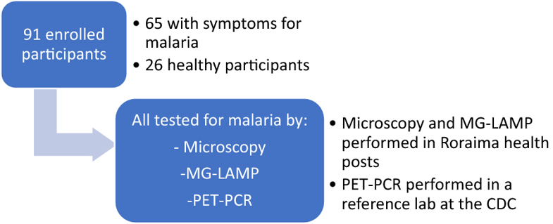
Fig. 1 Summary of enrolled patients and sample processing BACKGROUND: Microscopic detection of malaria parasites is the standard method for clinical diagnosis of malaria in Brazil. However, malaria epidemiological surveillance studies specifically aimed at the detection of low-density infection and asymptomatic cases will require more sensitive and field-usable tools. The diagnostic accuracy of the colorimetric …