Generic selectors
Exact matches only
Search in title
Search in content
Post Type Selectors

Tag: Kojo Mensa-Wilmot

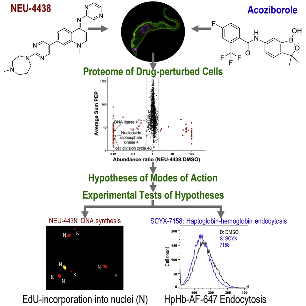
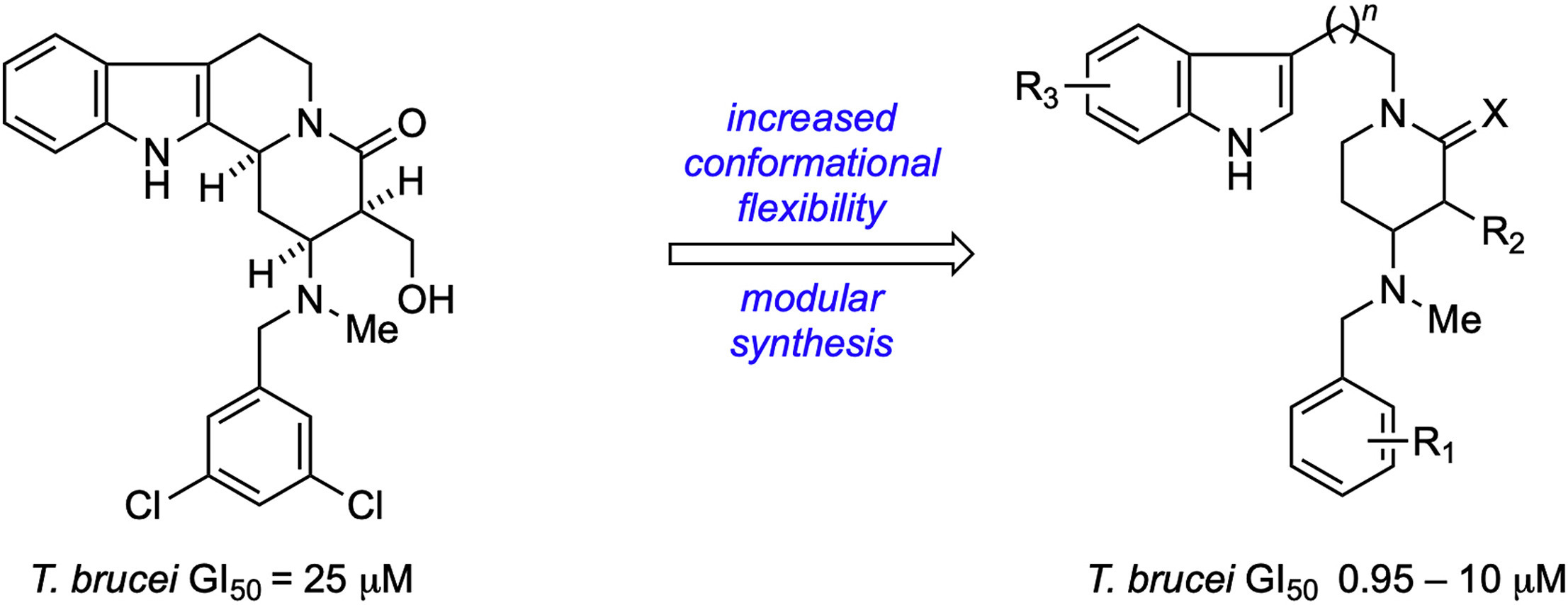
Chemical Optimization of CBL0137 for Human African Trypanosomiasis Lead Drug Discovery

The carbazole CBL0137 (1) is a lead for drug development against human African trypanosomiasis (HAT), a disease caused by Trypanosoma brucei. To advance 1 as a candidate drug, we synthesized new analogs that were evaluated for the physicochemical properties, antitrypanosome potency, selectivity against human cells, metabolism in microsomes or hepatocytes, and efflux ratios. Structure-activity/property analyses of analogs revealed eight new compounds with higher or equivalent selectivity indices (5j5t5v5w5y8d13i, and 22e). Based on the overall compound profiles, compounds 5v and 5w were selected for assessment in a mouse model of HAT; while 5v demonstrated a lead-like profile for HAT drug development, 5w showed a lack of efficacy. Lessons from these studies will inform further optimization of carbazoles for HAT and other indications.

Baljinder Singh, Amrita Sharma, Naresh Gunaganti, Mitch Rivers, Pradip K Gadekar, Brady Greene, Michael Chichioco, Carlos E Sanz-Rodriguez, Courtney Fu, Catherine LeBlanc, Erin Burchfield, Nyle Sharif, Benjamin Hoffman, Gaurav Kumar, Andrei Purmal, Kojo Mensa-Wilmot, Michael P Pollastri. J Med Chem. 2023 Jan 25. doi: 10.1021/acs.jmedchem.2c01767.

Pseudokinase NRP1 facilitates endocytosis of transferrin in the African trypanosome

Trypanosoma brucei causes human African trypanosomiasis (HAT) and nagana in cattle. During infection of a vertebrate, endocytosis of host transferrin (Tf) is important for viability of the parasite. The majority of proteins involved in trypanosome endocytosis of Tf are unknown. Here we identify pseudokinase NRP1 (Tb427tmp.160.4770) as a regulator of Tf endocytosis. Genetic knockdown of NRP1 inhibited endocytosis of Tf without blocking uptake of bovine serum albumin. Binding of Tf to the flagellar pocket was not affected by knockdown of NRP1. However the quantity of Tf per endosome dropped significantly, consistent with NRP1 promoting robust capture and/or retention of Tf in vesicles. NRP1 is involved in motility of Tf-laden vesicles since distances between endosomes and the kinetoplast were reduced after knockdown of the gene. In search of possible mediators of NRP1 modulation of Tf endocytosis, the gene was knocked down and the phosphoproteome analyzed. Phosphorylation of protein kinases forkhead, NEK6, and MAPK10 was altered, in addition to EpsinR, synaptobrevin and other vesicle-associated proteins predicted to be involved in endocytosis. These candidate proteins may link NRP1 functionally either to protein kinases or to vesicle-associated proteins.

Gaurav Kumar, Bryanna Thomas, Kojo Mensa-Wilmot. Sci Rep. 2022 Nov 3;12(1):18572. doi: 10.1038/s41598-022-22054-x.

Hypothesis-generating proteome perturbation to identify NEU-4438 and acoziborole modes of action in the African Trypanosome

NEU-4438 is a lead for the development of drugs against Trypanosoma brucei, which causes human African trypanosomiasis. Optimized with phenotypic screening, targets of NEU-4438 are unknown. Herein, we present a cell perturbome workflow that compares NEU-4438’s molecular modes of action to those of SCYX-7158 (acoziborole). Following a 6 h perturbation of trypanosomes, NEU-4438 and acoziborole reduced steady-state amounts of 68 and 92 unique proteins, respectively. After analysis of proteomes, hypotheses formulated for modes of action were tested: Acoziborole and NEU-4438 have different modes of action. Whereas NEU-4438 prevented DNA biosynthesis and basal body maturation, acoziborole destabilized CPSF3 and other proteins, inhibited polypeptide translation, and reduced endocytosis of haptoglobin-hemoglobin. These data point to CPSF3-independent modes of action for acoziborole. In case of polypharmacology, the cell-perturbome workflow elucidates modes of action because it is target-agnostic. Finally, the workflow can be used in any cell that is amenable to proteomic and molecular biology experiments.

Amrita Sharma, Michael Cipriano, Lori Ferrins, Stephen L Hajduk, Kojo Mensa-Wilmot. iScience. 2022 Oct 7;25(11):105302. doi: 10.1016/j.isci.2022.105302. eCollection 2022 Nov 18.

Casein kinase TbCK1.2 regulates division of kinetoplast DNA, and movement of basal bodies in the African trypanosome

The single mitochondrial nucleoid (kinetoplast) of Trypanosoma brucei is found proximal to a basal body (mature (mBB)/probasal body (pBB) pair). Kinetoplast inheritance requires synthesis of, and scission of kinetoplast DNA (kDNA) generating two kinetoplasts that segregate with basal bodies into daughter cells. Molecular details of kinetoplast scission and the extent to which basal body separation influences the process are unavailable. To address this topic, we followed basal body movements in bloodstream trypanosomes following depletion of protein kinase TbCK1.2 which promotes kinetoplast division. In control cells we found that pBBs are positioned 0.4 um from mBBs in G1, and they mature after separating from mBBs by at least 0.8 um: mBB separation reaches ~2.2 um. These data indicate that current models of basal body biogenesis in which pBBs mature in close proximity to mBBs may need to be revisited. Knockdown of TbCK1.2 produced trypanosomes containing one kinetoplast and two nuclei (1K2N), increased the percentage of cells with uncleaved kDNA 400%, decreased mBB spacing by 15%, and inhibited cytokinesis 300%. We conclude that (a) separation of mBBs beyond a threshold of 1.8 um correlates with division of kDNA, and (b) TbCK1.2 regulates kDNA scission. We propose a Kinetoplast Division Factor hypothesis that integrates these data into a pathway for biogenesis of two daughter mitochondrial nucleoids.

Catherine Sullenberger, Benjamin Hoffman, Justin Wiedeman, Gaurav Kumar, Kojo Mensa-Wilmot. PLoS One. 2021 Apr 16;16(4):e0249908. doi: 10.1371/journal.pone.0249908.

Design, Synthesis, and Evaluation of Novel Anti-Trypanosomal Compounds

Human African trypanosomiasis (HAT) is a deadly neglected tropical disease caused by the protozoan parasite Trypanosoma brucei. During the course of screening a collection of diverse nitrogenous heterocycles, we discovered two novel compounds that contain the tetracyclic core of the Yohimbine and Corynanthe alkaloids, were potent inhibitors of T. brucei proliferation and T. brucei methionyl-tRNA synthetase (TbMetRS) activity. Inspired by these key findings, we prepared several novel series of hydroxyalkyl δ-lactam, δ-lactam, and piperidine analogs and tested their anti-trypanosomal activity. A number of inhibitors are more potent against T. brucei than these initial hits with one hydroxyalkyl δ-lactam derivative being 25-fold more effective in our assay. Surprisingly, most of these active compounds failed to inhibit TbMetRS. This work underscores the importance of verifying, irrespective of close structural similarities, that new compounds designed from a lead with a known biological target engage the putative binding site.

Lance T. Lepovitz, Alan R. Meis, Sarah M. Thomas, Justin Wiedeman, Alexandra Pham, Kojo Mensa-Wilmot, Stephen F. Martin. Tetrahedron. 2020 Apr 17;76(16). pii: 131086. doi: 10.1016/j.tet.2020.131086

Kinetoplast Division Factors in a Trypanosome

Trypanosoma brucei

Highlights

  • Kinetoplasts (mitochondrial genome nucleoids) are important in bloodstream trypanosomes for the establishment of mitochondrial membrane potential.
  • Many proteins involved in segregation of kinetoplasts have been identified.
  • A region between a kinetoplast and basal bodies is described as a tripartite attachment complex (TAC).
  • A set of TAC-associated proteins (TACAPs) has been proposed as the machinery for kinetoplast segregation.
  • Subcomplexes of TACAPs that form in vivo have been described.
  • Several proteins that do not associate with TAC are involved in the maintenance of the kinetoplast.
  • New kinetoplast-associated proteins have been identified.
  • We are approaching an exciting period in the field when a molecular understanding of how all aspects of kinetoplast biogenesis are executed seems achievable.

Kojo Mensa-Wilmot, Benjamin Hoffman, Justin Wiedman, Catherine Sullenberger, Amrita Sharma. 2019. Trends in Parasitology. https://doi.org/10.1016/j.pt.2018.11.002

Anilinoquinoline based inhibitors of trypanosomatid proliferation

Abstract

We recently reported the medicinal chemistry re-optimization of a series of compounds derived from the human tyrosine kinase inhibitor, lapatinib, for activity against Plasmodium falciparum. From this same library of compounds, we now report potent compounds against Trypanosoma brucei brucei (which causes human African trypanosomiasis), Tcruzi (the pathogen that causes Chagas disease), and Leishmania spp. (which cause leishmaniasis). In addition, sub-micromolar compounds were identified that inhibit proliferation of the parasites that cause African animal trypanosomiasis, Tcongolense and Tvivax. We have found that this set of compounds display acceptable physicochemical properties and represent progress towards identification of lead compounds to combat several neglected tropical diseases.

Lori Ferrins, Amrita Sharma, Sarah M. Thomas, Naimee Mehta, Jessey Erath, Scott Tanghe, Susan E. Leed, Ana Rodriguez, Kojo Mensa-Wilmot, Richard J. Sciotti, Kirsten Gillingwater, Michael P. Pollastri. 2018. PLOS Neglected Tropical Diseases. https://doi.org/10.1371/journal.pntd.0006834

Series of Alkynyl-Substituted Thienopyrimidines as Inhibitors of Protozoan Parasite Proliferation

graphical abstract

Abstract

Discovery of new chemotherapeutic lead agents can be accelerated by optimizing chemotypes proven to be effective in other diseases to act against parasites. One such medicinal chemistry campaign has focused on optimizing the anilinoquinazoline drug lapatinib (1) and the alkynyl thieno[3,2-d]pyrimidine hit GW837016X (NEU-391, 3) into leads for antitrypanosome drugs. We now report the structure–activity relationship studies of 3 and its analogs against Trypanosoma brucei, which causes human African trypanosomiasis (HAT). The series was also tested against Trypanosoma cruziLeishmania major, and Plasmodium falciparum. In each case, potent antiparasitic hits with acceptable toxicity margins over mammalian HepG2 and NIH3T3 cell lines were identified. In a mouse model of HAT, 3 extended life of treated mice by 50%, compared to untreated controls. At the cellular level, 3 inhibited mitosis and cytokinesis in T. brucei. Thus, the alkynylthieno[3,2-d]pyrimidine chemotype is an advanced hit worthy of further optimization as a potential chemotherapeutic agent for HAT.

Jennifer L. Woodring, Ranjan Behera, Amrita Sharma, Justin Wiedeman, Gautam Patel, Baljinder Singh, Paul Guyett, Emanuele Amata, Jessey Erath, Norma Roncal, Erica Penn, Susan E. Leed, Ana Rodriguez, Richard J. Sciotti, Kojo Mensa-Wilmot, and Michael P. Pollastri. 2018. ACS Med. Chem. Lett.; 9(10):996-1001. DOI: 10.1021/acsmedchemlett.8b00245

Trainee Adds New Tool to the Trypanosome Toolbox

Trypanosoma brucei stained with mCLING
mCLING-staining of membrane (green) and DAPI staining of DNA (magenta) in trypanosomes in various life cycle stages. Flagella appear as tube-like structures along the length of the cell body. Nanotubes can be seen projecting outward from the periphery of the cells.

When Ph.D. trainee Justin Wiedeman started investigating the role of protein kinase TbCK1.2, an enzyme found near the flagellum of Trypanosoma brucei, he quickly ran into a problem common to parasitologists. He needed a better tool for visualizing the membranes of this parasite. Since none of the membrane probes on the market quite did the job, he looked at how he could modify one for his purpose. He found a successful candidate in Synaptic Systems’ mCLING.

What is Trypanosoma brucei?

Trypanosoma brucei is a single cell parasite that causes Human African Trypanosomiasis (HAT), which is also known as African sleeping sickness. HAT occurs in 36 sub-Saharan countries where tsetse flies transmit the parasite to people and livestock. In cattle, the disease is known as nagana. Tsetse fly control efforts have drastically reduced the number of cases. According to the World Health Organization, in 2015, there were around 2,800 cases. However, a person can be infected for months or even years without symptoms. By the time symptoms become evident, the person is in the advanced stages of the disease and their central nervous system is impaired.

New tools are needed to study trypanosomes

There is still much to be learned about the parasite that could lead to better detection and more effective treatment. A major obstacle to the study of this tiny organism is the lack of tools and technology. Kojo Mensa-Wilmot’s research group in the Center for Tropical and Emerging Global Diseases at The University of Georgia has been instrumental in developing techniques and tools to increase the research community’s understanding of T. brucei. Now, Wiedeman has added a new tool to the trypanosome biology toolbox – a general method of outlining trypanosomes in fluorescence microscopy experiments.

“We are the first group to solve this general problem in super-resolution microscopy of T. brucei,” said Wiedeman. “mCLING is a highly versatile tool for studying trypanosome biology – it can be used with live or fixed trypanosomes.”

Fluorescent microscopy has been a leading method of studying T. brucei; however, there are limitations to this technology. Super-resolution microscopy offers great advantages over standard fluorescence microscopy. By employing several techniques to increase resolution, it allows for the observation of objects smaller than what can be seen with visible light.  Yet, it is not without its own limitations, most notably the inability to determine the periphery of cells. Without knowing the outer edges of the parasite, orientation of organelles and other structures within the cell is difficult.

“For Trypanosoma brucei, most of the membrane probes available do not work well in fixed trypanosomes,” said Wiedeman. “Researchers have been forced to use crude methods to outline trypanosomes in fluorescence microscopy.”

These “crude methods” include superimposing a transmitted light image or hand-drawing the outline. However, this workaround only allows for a two-dimensional study of the cell. Therefore, Wiedeman turned to a dye called mCLING that has been developed to track the membranes of neurons using super-resolution microscopy to see if he could adapt the technology to T. brucei membranes.

mCLING allows for the visualization of T. brucei membranes

“mCLING labels the flagellum and plasma membrane vividly, sometimes providing details of cell structure that rivals images obtained with scanning electron microscopy,” said Wiedeman.

Using a combination of standard-resolution and super-resolution fluorescence microscopy, he was able to confirm mCLING labels the plasma and flagellar membranes of T. brucei. Furthermore, using the Zeiss ELYRA S1 super-resolution microscopy in the Biomedical Microscopy Core, mCLING allowed for a 3D reconstruction of the parasite. This is the first time such an image has been reported. Finally, using the new ImageStream X Mark II in the CTEGD Cytometry Shared Resource Laboratory, he discovered mCLING could be used to track endocytosis (the process of importing molecules into the cell) in real time.

Recognizing mCLING’s potential to inform other studies of trypanosome biology, Weideman optimized protocols for using it with immunofluorescence assays and thus making possible what had been impossible with the overlay technique – visualizing the location of organelles in the vertical dimension relative to the cell body.

“It is especially well-suited for studying flagellar membrane biogenesis as well as kinetically tracking uptake of the plasma membrane into vesicles inside trypanosomes,” said Wiedeman. Other laboratories have already implemented these protocols in their own research. Steve Hajduk’s group, also in the Center for Tropical and Emerging Global Diseases, is using mCLING to study nanotubes in T. brucei.

This tool will allow for the study of trypanosomes in finer detail than ever before and the Mensa-Wilmot Research Group anticipates unlocking previously unseen secrets in T. brucei.


The full published study is available online: Wiedeman J, Mensa-Wilmot K (2018). A fixable probe for visualizing flagella and plasma membranes of the African trypanosome. PLoS One 13(5):e0197541. https://doi.org/10.1371/journal.pone.0197541

New UGA Drug Discovery Core lab works to develop treatment of leading diseases

Drug Discovery Center team
Members of the Drug Discovery Core steering committee in the new DDC laboratory (from left to right): Shelley Hooks, interim director of the Center for Drug Discovery and associate professor of pharmaceutical and biomedical sciences; Scott Pegan, chair of the steering committee and associate professor of pharmaceutical and biomedical sciences; Belen Cassera, associate professor of biochemistry and molecular biology; Kojo Mensa-Wilmot, professor and head of UGA’s cellular biology department and director of the Chemical Biology Group; and Brian Cummings, director of the Interdisciplinary Toxicology Program and professor of pharmaceutical and biomedical sciences.

Athens, Ga. – The University of Georgia has created the Drug Discovery Core laboratory, a campus-wide collaborative facility designed to hasten the development of therapeutic drugs for a number of major diseases.

A survey distributed to UGA researchers in 2016 identified chemical screening and toxicity profiling as the most critical needs for enhancing drug discovery research at UGA, and the DDC will address many of those needs for faculty working in infectious disease, regenerative medicine, cancer biology and other human health-focused disciplines.

Phase one of the new lab will allow for the curation, management and distribution of chemical libraries containing more than 50,000 compounds. The lab also will enable researchers to rapidly screen these chemical libraries in miniaturized models of various diseases using robotics and high-throughput signal detection. Finally, the lab will provide opportunities to identify potential toxicity of the compounds and determine if their chemical properties will allow them to be successfully delivered to patients. Additional capabilities, including pharmacokinetic characterization, genotoxicity and assay design, are under development.

“The most immediate outcome of the DDC lab will be to generate preliminary data from pilot chemical screens, which is necessary to secure large drug discovery grants from the National Institutes of Health to fund more advanced drug discovery research,” said Shelley Hooks, interim director of the Center for Drug Discovery and associate professor of pharmaceutical and biomedical science. “The longer-term goals of the lab are to discover and develop new drug candidates and chemical probes, as well as enhance training of graduate students in biotechnology.”

Creation of the DDC was initiated by Hooks in collaboration with Brian Cummings, director of the Interdisciplinary Toxicology Program and professor in the pharmaceutical and biomedical sciences department, and Scott Pegan, chair of the DDC steering committee and associate professor of pharmaceutical and biomedical sciences.

Sponsoring campus organizations include the College of Pharmacy, the College of Veterinary Medicine, the Office of Research, the Center for Tropical and Emerging Diseases and the Department of Cellular Biology.

The laboratory is located in Room 224 of the Wilson Building in the College of Pharmacy. For more information on capability, resources and access to the libraries and screening instruments, contact Pegan (spegan@uga.edu) or see cdd.rx.uga.edu.

Writer: Mickey Y. Montevideo
Contact: Shelley B. Hooks