Generic selectors
Exact matches only
Search in title
Search in content
Post Type Selectors

Category: publications

Massive invasion of organellar DNA drives nuclear genome evolution in Toxoplasma
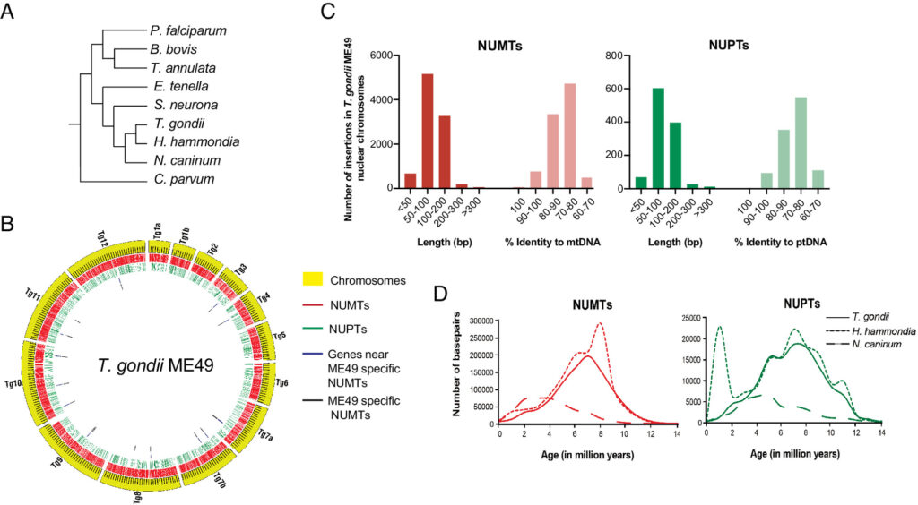
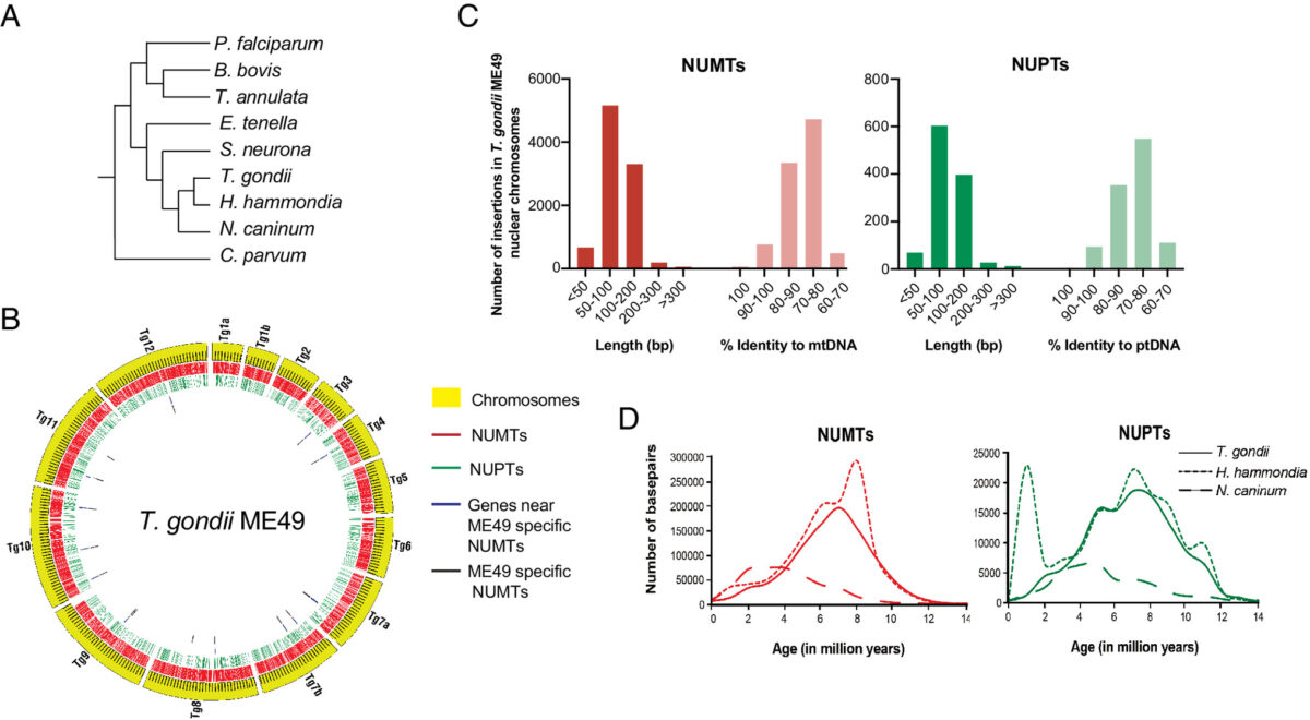
Figure 1 Characteristics of NUMTs and NUPTs in T. gondii ME49
Fig. 1 Characteristics of NUMTs and NUPTs in T. gondii ME49


Toxoplasma gondii is a zoonotic protist pathogen that infects up to one third of the human population. This apicomplexan parasite contains three genome sequences: nuclear (65 Mb); plastid organellar, ptDNA (35 kb); and mitochondrial organellar, mtDNA (5.9 kb of non-repetitive sequence). We find that the nuclear genome contains a significant amount of NUMTs (nuclear integrants of mitochondrial DNA) and NUPTs (nuclear integrants of plastid DNA) that are continuously acquired and represent a significant source of intraspecific genetic variation. NUOT (nuclear DNA of organellar origin) accretion has generated 1.6% of the extant T. gondii ME49 nuclear genome-the highest fraction ever reported in any organism. NUOTs are primarily found in organisms that retain the non-homologous end-joining repair pathway. Significant movement of organellar DNA was experimentally captured via amplicon sequencing of a CRISPR-induced double-strand break in non-homologous end-joining repair competent, but not ku80 mutant, Toxoplasma parasites. Comparisons with Neospora caninum, a species that diverged from Toxoplasma ~28 mya, revealed that the movement and fixation of five NUMTs predates the split of the two genera. This unexpected level of NUMT conservation suggests evolutionary constraint for cellular function. Most NUMT insertions reside within (60%) or nearby genes (23% within 1.5 kb), and reporter assays indicate that some NUMTs have the ability to function as cis-regulatory elements modulating gene expression. Together, these findings portray a role for organellar sequence insertion in dynamically shaping the genomic architecture and likely contributing to adaptation and phenotypic changes in this important human pathogen.

Sivaranjani Namasivayam, Cheng Sun, Assiatu B Bah, Jenna Oberstaller, Edwin Pierre-Louis, Ronald Drew Etheridge, Cedric Feschotte, Ellen J Pritham, Jessica C Kissinger. Proc Natl Acad Sci U S A. 2023 Nov 7;120(45):e2308569120. doi: 10.1073/pnas.2308569120.

Insulin-like peptides and ovary ecdysteroidogenic hormone differentially stimulate physiological processes regulating egg formation in the mosquito Aedes aegypti

graphical abstract

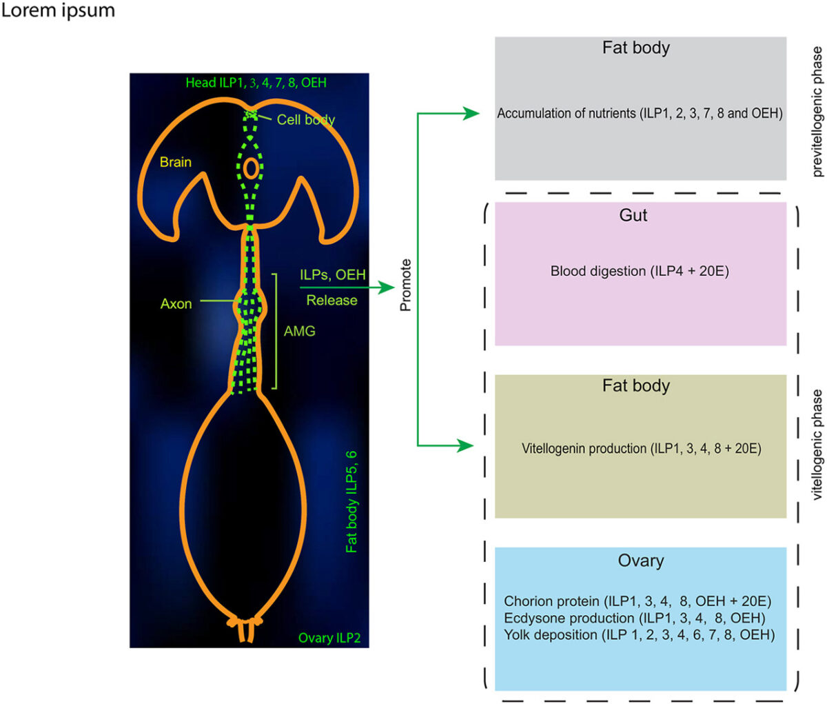
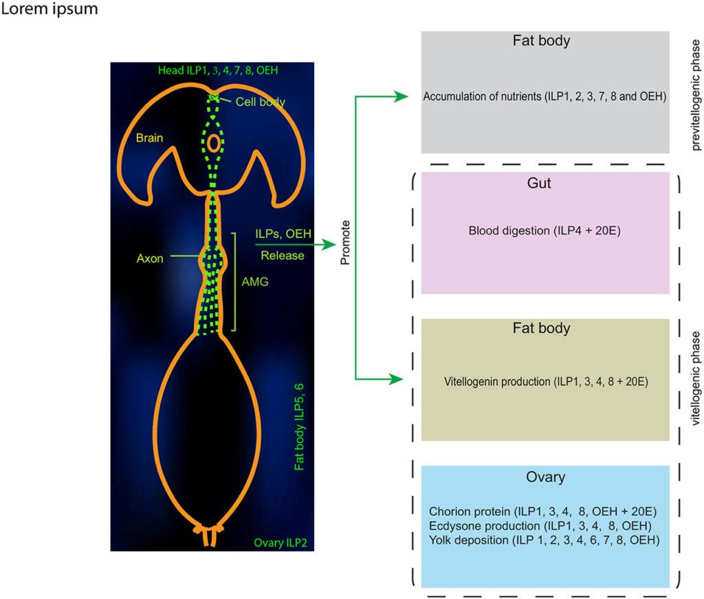
Mosquitoes including Aedes aegypti are human disease vectors because females must blood feed to produce and lay eggs. Blood feeding triggers insulin-insulin growth factor signaling (IIS) which regulates several physiological processes required for egg development. A. aegypti encodes 8 insulin-like peptides (ILPs) and one insulin-like receptor (IR) plus ovary ecdysteroidogenic hormone (OEH) that also activates IIS through the OEH receptor (OEHR). In this study, we assessed the expression of A. aegypti ILPs and OEH during a gonadotropic cycle and produced each that were functionally characterized to further understand their roles in regulating egg formation. All A. aegypti ILPs and OEH were expressed during a gonadotropic cycle. Five ILPs (1, 3, 4, 7, 8) and OEH were specifically expressed in the head, while antibodies to ILP3 and OEH indicated each was released after blood feeding from ventricular axons that terminate on the anterior midgut. A subset of ILP family members and OEH stimulated nutrient storage in previtellogenic females before blood feeding, whereas most IIS-dependent processes after blood feeding were activated by one or more of the brain-specific ILPs and/or OEH. ILPs and OEH with different biological activities also exhibited differences in IIS as measured by phosphorylation of the IR, phosphoinositide 3-kinase/Akt kinase (AKT) and mitogen-activated protein kinase/extracellular signal-regulated kinase (ERK). Altogether, our results provide the first results that compare the functional activities of all ILP family members and OEH produced by an insect.

Kangkang Chen, Xiaoyi Dou, Jai Hoon Eum, Ruby A Harrison, Mark R Brown, Michael R Strand. Insect Biochem Mol Biol. 2023 Oct 30:104028. doi: 10.1016/j.ibmb.2023.104028.

Functional characterization of Microplitis demolitor bracovirus genes that encode nucleocapsid

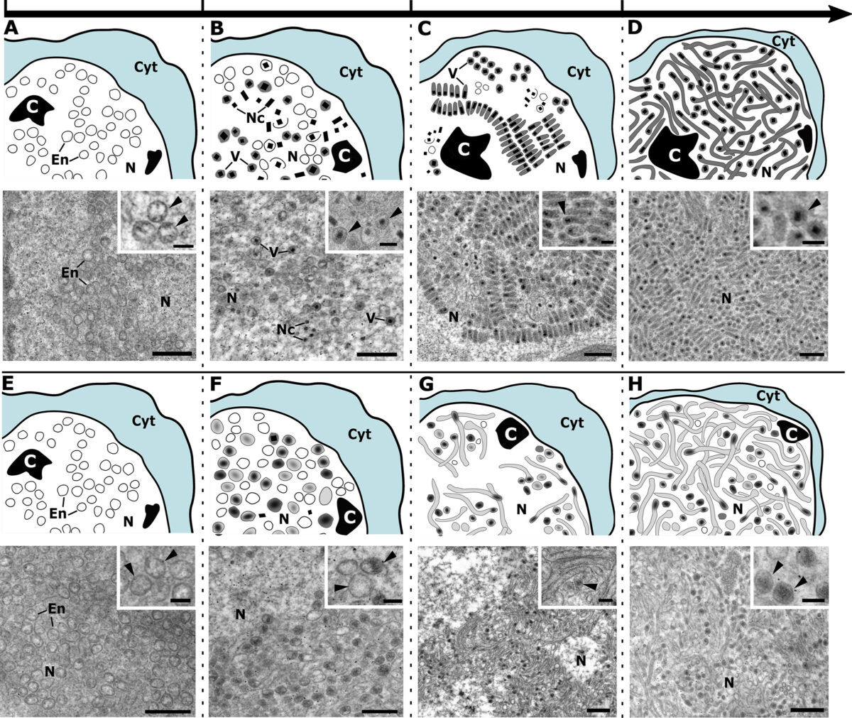
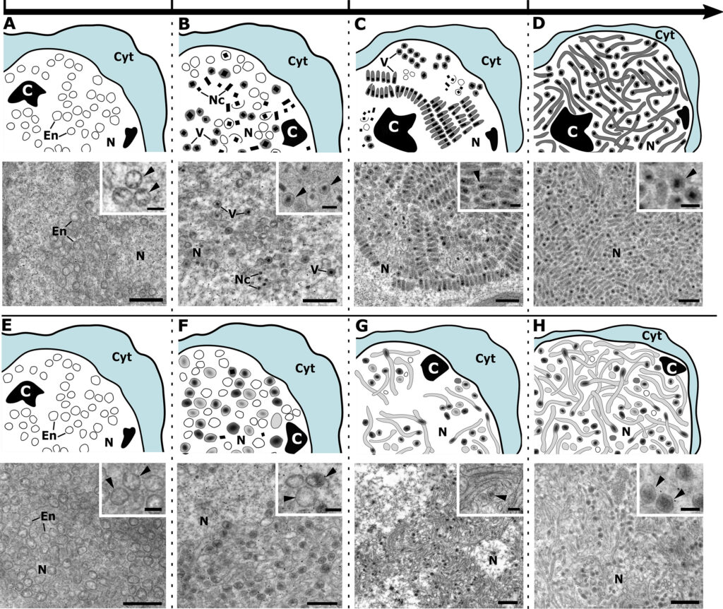
Fig 2 Virion morphogenesis (phases 1–4) in calyx cells from newly emerged adult females injected with ds-eGFP (control) (A–D) or ds-vp39 (E–H).


Bracoviruses (BVs) are endogenized nudiviruses in parasitoid wasps of the microgastroid complex (order Hymenoptera: Family Braconidae). BVs produce replication-defective virions that adult female wasps use to transfer DNAs encoding virulence genes to parasitized hosts. Some BV genes are shared with nudiviruses and baculoviruses with studies of the latter providing insights on function, whereas other genes are only known from nudiviruses or other BVs which provide no functional insights. A proteomic analysis of Microplitis demolitor bracovirus (MdBV) virions recently identified 16 genes encoding nucleocapsid components. In this study, we further characterized most of these genes. Some nucleocapsid genes exhibited early or intermediate expression profiles, while others exhibited late expression profiles. RNA interference (RNAi) assays together with transmission electron microscopy indicated vp39HzNVorf9-like2HzNVorf93-likeHzNVorf106-likeHzNVorf118-likeand 27b are required to produce capsids with a normal barrel-shaped morphology. RNAi knockdown of vlf-1avlf-1b-1vlf-1b-2int-1, and p6.9-1 did not alter the formation of barrel-shaped capsids but each reduced processing of amplified proviral segments and DNA packaging as evidenced by the formation of electron translucent capsids. All of the genes required for normal capsid assembly were also required for proviral segment processing and DNA packaging. Collectively, our results deorphanize several BV genes with previously unknown roles in virion morphogenesis.IMPORTANCEUnderstanding how bracoviruses (BVs) function in wasps is of broad interest in the study of virus evolution. This study characterizes most of the Microplitis demolitor bracovirus (MdBV) genes whose products are nucleocapsid components. Results indicate several genes unknown outside of nudiviruses and BVs are essential for normal capsid assembly. Results also indicate most MdBV tyrosine recombinase family members and the DNA binding protein p6.9-1 are required for DNA processing and packaging into nucleocapsids.

Ange Lorenzi, Michael J Arvin, Gaelen R Burke, Michael R Strand. J Virol. 2023 Oct 25:e0081723. doi: 10.1128/jvi.00817-23.

Shotgun Kinetic Target-Guided Synthesis Approach Enables the Discovery of Small-Molecule Inhibitors against Pathogenic Free-Living Amoeba Glucokinases

Pathogenic free-living amoebae (pFLA) can cause life-threatening central nervous system (CNS) infections and warrant the investigation of new chemical agents to combat the rise of infection from these pathogens. Naegleria fowleri glucokinase (NfGlck), a key metabolic enzyme involved in generating glucose-6-phosphate, was previously identified as a potential target due to its limited sequence similarity with human Glck (HsGlck). Herein, we used our previously demonstrated multifragment kinetic target-guided synthesis (KTGS) screening strategy to identify inhibitors against pFLA glucokinases. Unlike the majority of previous KTGS reports, our current study implements a “shotgun” approach, where fragments were not biased by predetermined binding potentials. The study resulted in the identification of 12 inhibitors against 3 pFLA glucokinase enzymes─NfGlck, Balamuthia mandrillaris Glck (BmGlck), and Acanthamoeba castellanii Glck (AcGlck). This work demonstrates the utility of KTGS to identify small-molecule binders for biological targets where resolved X-ray crystal structures are not readily accessible.

Mintesinot Kassu, Prakash T Parvatkar, Jillian Milanes, Neil P Monaghan, Chungsik Kim, Matthew Dowgiallo, Yingzhao Zhao, Ami H Asakawa, Lili Huang, Alicia Wagner, Brandon Miller, Karissa Carter, Kayleigh F Barrett, Logan M Tillery, Lynn K Barrett, Isabelle Q Phan, Sandhya Subramanian, Peter J Myler, Wesley C Van Voorhis, James W Leahy, Christopher A Rice, Dennis E Kyle, James Morris, Roman Manetsch. ACS Infect Dis. 2023 Oct 11. doi: 10.1021/acsinfecdis.3c00284.

ATP synthase-associated coiled-coil-helix-coiled-coil-helix (CHCH) domain-containing proteins are critical for mitochondrial function in Toxoplasma gondii

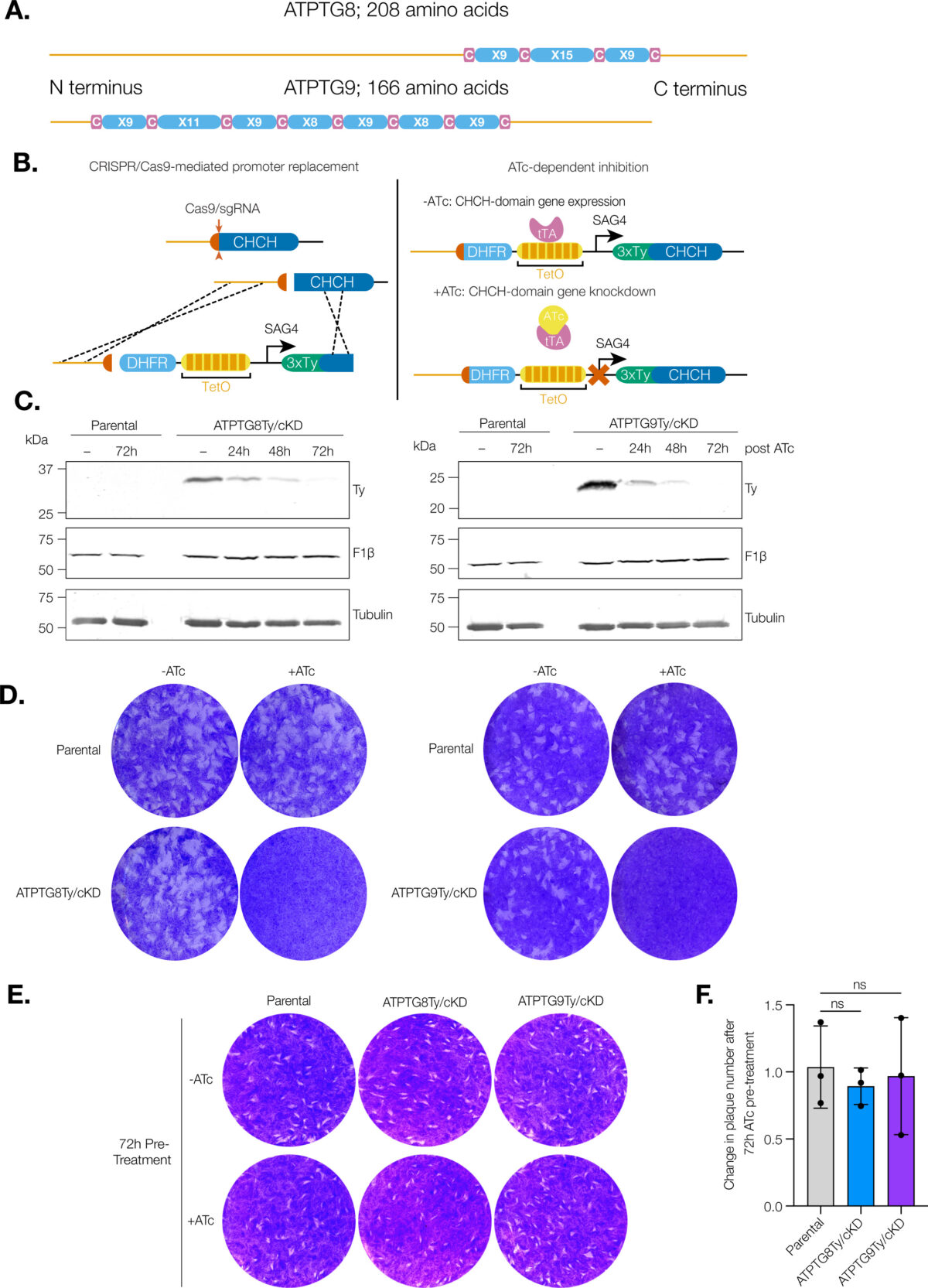
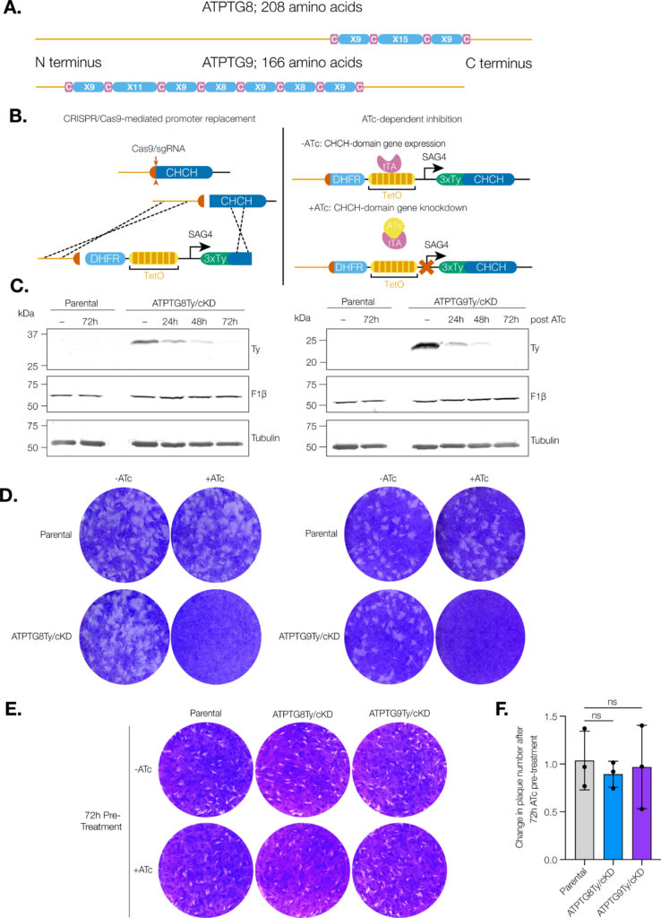
CHCH domain proteins associated with the T. gondii ATP synthase are essential for the lytic cycle. (A) Schematic representation of the CHCH domain size and location in ATPTG8 and ATPTG9. C represents cysteine residues and X represents any other amino acid residue.

Coiled-coil-helix-coiled-coil-helix (CHCH) domains consist of two pairs of cysteine residues that are oxidized to form disulfide bonds upon mitochondrial import. Proteins containing these domains play important roles in mitochondrial ultrastructure and in the biogenesis, function, and stability of electron transport chain complexes. Interestingly, recent investigations of the Toxoplasma gondii ATP synthase identified subunits containing CHCH domains. As CHCH domain proteins have never been found in any other ATP synthase, their role in T. gondii was unclear. Using conditional gene knockdown systems, we investigated two T. gondii ATP synthase subunits containing CHCH domains: ATPTG8 and ATPTG9. We show that these two subunits are essential for the lytic cycle as well as stability and function of the ATP synthase. Further, we illustrated that their knockdown disrupts multiple aspects of mitochondrial morphology, including ultrastructure and cristae density. Mutation of key cysteine residues in the CHCH domains also caused mis-localization of the proteins. Our work suggests that these proteins likely provide structural support to the exceptionally large T. gondii ATP synthase complex and that perturbations to the structural integrity of this complex result in deleterious downstream effects on the parasite mitochondrion. These investigations add to a growing body of work focused on the divergent aspects of the apicomplexan ATP synthase, which could ultimately uncover novel drug targets. IMPORTANCE Members of the coiled-coil-helix-coiled-coil-helix (CHCH) domain protein family are transported into the mitochondrial intermembrane space, where they play important roles in the biogenesis and function of the organelle. Unexpectedly, the ATP synthase of the apicomplexan Toxoplasma gondii harbors CHCH domain-containing subunits of unknown function. As no other ATP synthase studied to date contains this class of proteins, characterizing their function will be of broad interest to the fields of molecular parasitology and mitochondrial evolution. Here, we demonstrate that that two T. gondii ATP synthase subunits containing CHCH domains are required for parasite survival and for stability and function of the ATP synthase. We also show that knockdown disrupts multiple aspects of the mitochondrial morphology of T. gondii and that mutation of key residues in the CHCH domains caused mis-localization of the proteins. This work provides insight into the unique features of the apicomplexan ATP synthase, which could help to develop therapeutic interventions against this parasite and other apicomplexans, such as the malaria-causing parasite Plasmodium falciparum.

Madelaine M Usey, Diego Huet. mBio. 2023 Oct 5:e0176923. doi: 10.1128/mbio.01769-23.

Time-resolved proximity biotinylation implicates a porin protein in export of transmembrane malaria parasite effectors

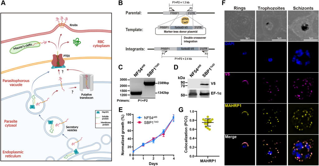
Figure 1 Generation of SBP1TbID mutants.
Generation of SBP1TbID mutants.

The malaria-causing parasite, Plasmodium falciparum completely remodels its host red blood cell (RBC) through the export of several hundred parasite proteins, including transmembrane proteins, across multiple membranes to the RBC. However, the process by which these exported membrane proteins are extracted from the parasite plasma membrane for export remains unknown. To address this question, we fused the exported membrane protein, skeleton binding protein 1 (SBP1), with TurboID, a rapid, efficient, and promiscuous biotin ligase (SBP1TbID). Using time-resolved, proximity biotinylation, and label-free quantitative proteomics, we identified two groups of SBP1TbID interactors: early interactors (pre-export) and late interactors (post-export). Notably, two promising membrane-associated proteins were identified as pre-export interactors, one of which possesses a predicted translocon domain, that could facilitate the export of membrane proteins. Further investigation using conditional mutants of these candidate proteins showed that these proteins were essential for asexual growth and localize to the host-parasite interface during early stages of the intraerythrocytic cycle. These data suggest that they may play a role in ushering membrane proteins from the PPM for export to the host RBC.

David Anaguano, Watcharatip Dedkhad, Carrie F Brooks, David W Cobb, Vasant Muralidharan. J Cell Sci. 2023 Sep 29;jcs.260506. doi: 10.1242/jcs.260506

Characterization of the extracellular vesicles, ultrastructural morphology, and intercellular interactions of multiple clinical isolates of the brain-eating amoeba, Naegleria fowleri

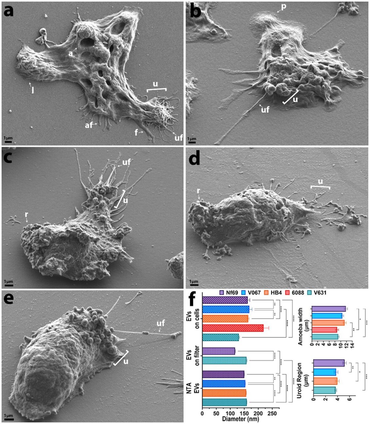
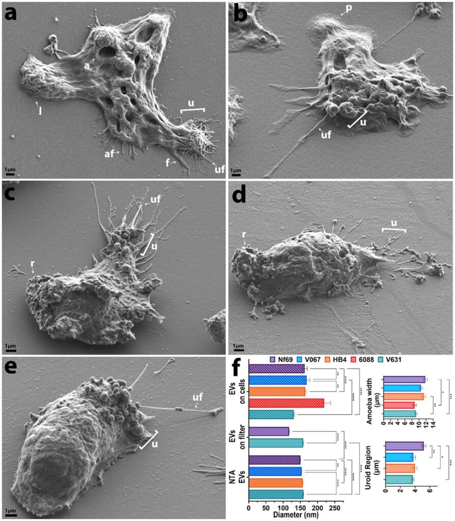
SEM micrographs of each clinical isolate in axenic culture.

Introduction: As global temperatures rise to unprecedented historic levels, so too do the latitudes of habitable niches for the pathogenic free-living amoeba, Naegleria fowleri. This opportunistic parasite causes a rare, but >97% fatal, neurological infection called primary amoebic meningoencephalitis. Despite its lethality, this parasite remains one of the most neglected and understudied parasitic protozoans.

Methods: To better understand amoeboid intercellular communication, we elucidate the structure, proteome, and potential secretion mechanisms of amoeba-derived extracellular vesicles (EVs), which are membrane-bound communication apparatuses that relay messages and can be used as biomarkers for diagnostics in various diseases.

Results and discussion: Herein we propose that N. fowleri secretes EVs in clusters from the plasma membrane, from multivesicular bodies, and via beading of thin filaments extruding from the membrane. Uptake assays demonstrate that EVs are taken up by other amoebae and mammalian cells, and we observed a real-time increase in metabolic activity for mammalian cells exposed to EVs from amoebae. Proteomic analysis revealed >2,000 proteins within the N. fowleri-secreted EVs, providing targets for the development of diagnostics or therapeutics. Our work expands the knowledge of intercellular interactions among these amoebae and subsequently deepens the understanding of the mechanistic basis of PAM.

A Cassiopeia Russell, Peter Bush, Gabriela Grigorean, Dennis E Kyle. Front Microbiol. 2023 Sep 27:14:1264348. doi: 10.3389/fmicb.2023.1264348.

Sheptide A: an antimalarial cyclic pentapeptide from a fungal strain in the Herpotrichiellaceae

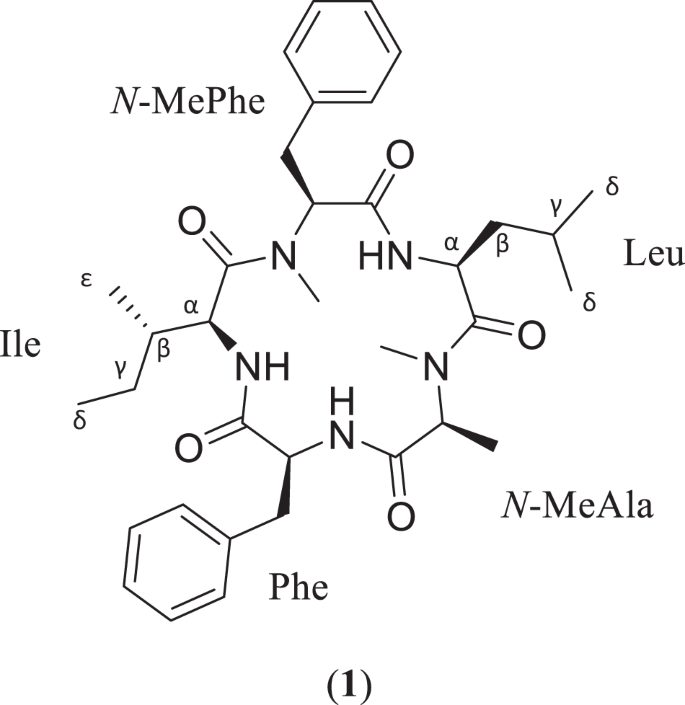
Structure and amino acid sequence of the cyclic pentapeptide, sheptide A (1)

As part of ongoing efforts to isolate biologically active fungal metabolites, a cyclic pentapeptide, sheptide A (1), was discovered from strain MSX53339 (Herpotrichiellaceae). The structure and sequence of 1 were determined primarily by analysis of 2D NMR and HRMS/MS data, while the absolute configuration was assigned using a modified version of Marfey’s method. In an in vitro assay for antimalarial potency, 1 displayed a pEC50 value of 5.75 ± 0.49 against malaria-causing Plasmodium falciparum. Compound 1 was also tested in a counter screen for general cytotoxicity against human hepatocellular carcinoma (HepG2), yielding a pCC50 value of 5.01 ± 0.45 and indicating a selectivity factor of ~6. This makes 1 the third known cyclic pentapeptide biosynthesized by fungi with antimalarial activity.

Robert A Shepherd, Cody E Earp, Kristof B Cank, Huzefa A Raja, Joanna Burdette, Steven P Maher, Adriana A Marin, Anthony A Ruberto, Sarah Lee Mai, Blaise A Darveaux, Dennis E Kyle, Cedric J Pearce, Nicholas H Oberlies. J Antibiot (Tokyo). 2023 Sep 20. doi: 10.1038/s41429-023-00655-6.

Synergy between a cytoplasmic vWFA/VIT protein and a WD40-repeat F-box protein controls development in Dictyostelium

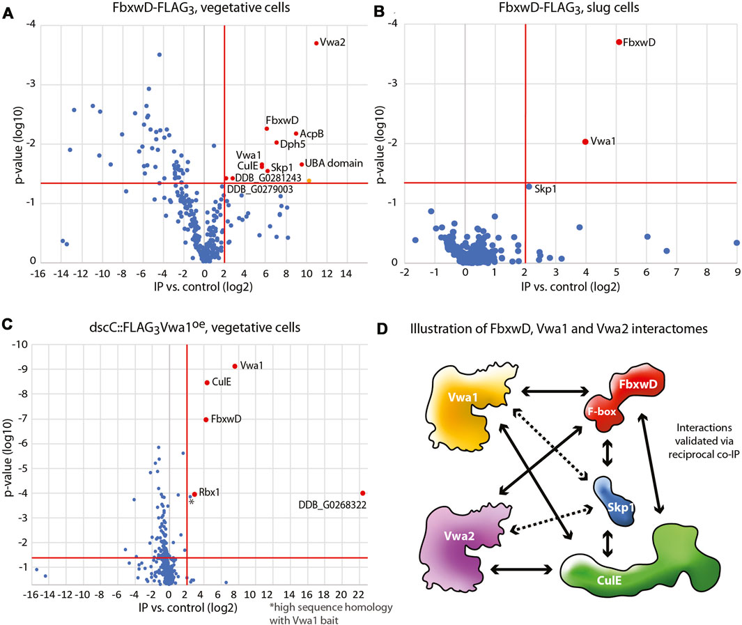
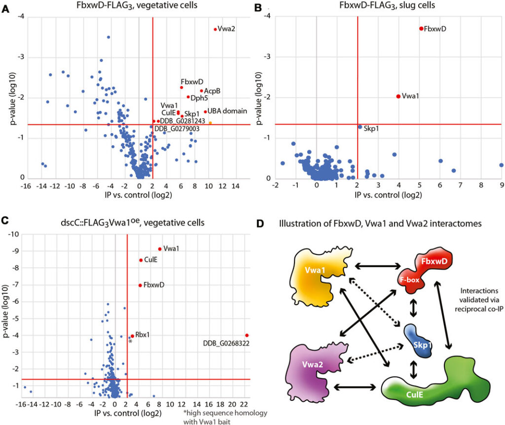
Interactomes of FbxwD-FLAG3 and FLAG3Vwa1. Immunoprecipitations of FLAG tagged targets using anti-FLAG mAb M2 from cells solubilized in non-ionic detergent (0.2% NP-40) were subjected to a proteomics work-flow that included generation of peptides with endo Lys-C and trypsin followed by detection by nLC MS/MS and quantitation by spectral counting.

Like most eukaryotes, the pre-metazoan social amoeba Dictyostelium depends on the SCF (Skp1/cullin-1/F-box protein) family of E3 ubiquitin ligases to regulate its proteome. In Dictyostelium, starvation induces a transition from unicellular feeding to a multicellular slug that responds to external signals to culminate into a fruiting body containing terminally differentiated stalk and spore cells. These transitions are subject to regulation by F-box proteins and O2-dependent posttranslational modifications of Skp1. Here we examine in greater depth the essential role of FbxwD and Vwa1, an intracellular vault protein inter-alpha-trypsin (VIT) and von Willebrand factor-A (vWFA) domain containing protein that was found in the FbxwD interactome by co-immunoprecipitation. Reciprocal co-IPs using gene-tagged strains confirmed the interaction and similar changes in protein levels during multicellular development suggested co-functioning. FbxwD overexpression and proteasome inhibitors did not affect Vwa1 levels suggesting a non-substrate relationship. Forced FbxwD overexpression in slug tip cells where it is normally enriched interfered with terminal cell differentiation by a mechanism that depended on its F-box and RING domains, and on Vwa1 expression itself. Whereas vwa1-disruption alone did not affect development, overexpression of either of its three conserved domains arrested development but the effect depended on Vwa1 expression. Based on structure predictions, we propose that the Vwa1 domains exert their negative effect by artificially activating Vwa1 from an autoinhibited state, which in turn imbalances its synergistic function with FbxwD. Autoinhibition or homodimerization might be relevant to the poorly understood tumor suppressor role of the evolutionarily related VWA5A/BCSC-1 in humans.

Andrew W Boland, Elisabet Gas-Pascual, Hanke van der Wel, Hyun W Kim, Christopher M West. Front Cell Dev Biol. 2023 Sep 14;11:1259844. doi: 10.3389/fcell.2023.1259844. eCollection 2023.

Validation of a multiplex microsphere immunoassay for detection of antibodies to Trypanosoma cruzi in dogs

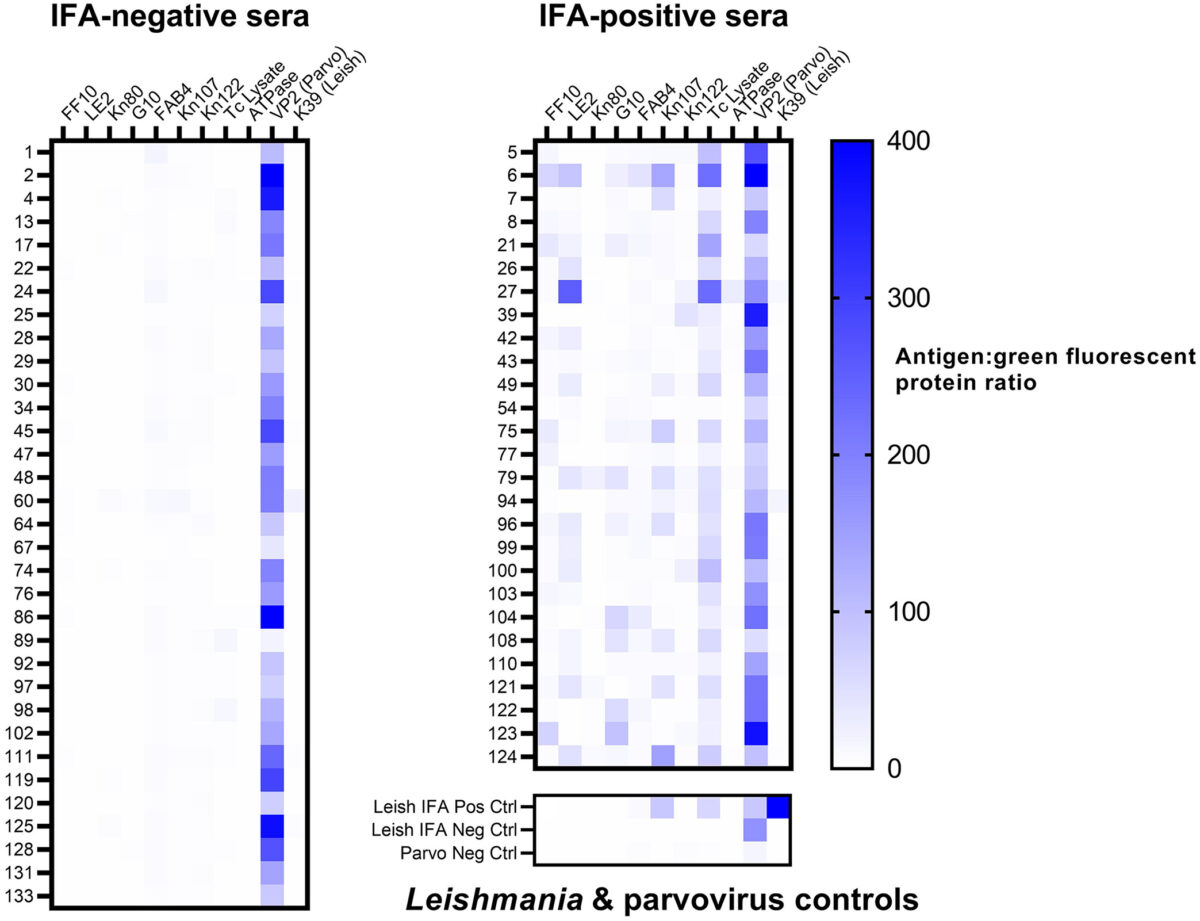
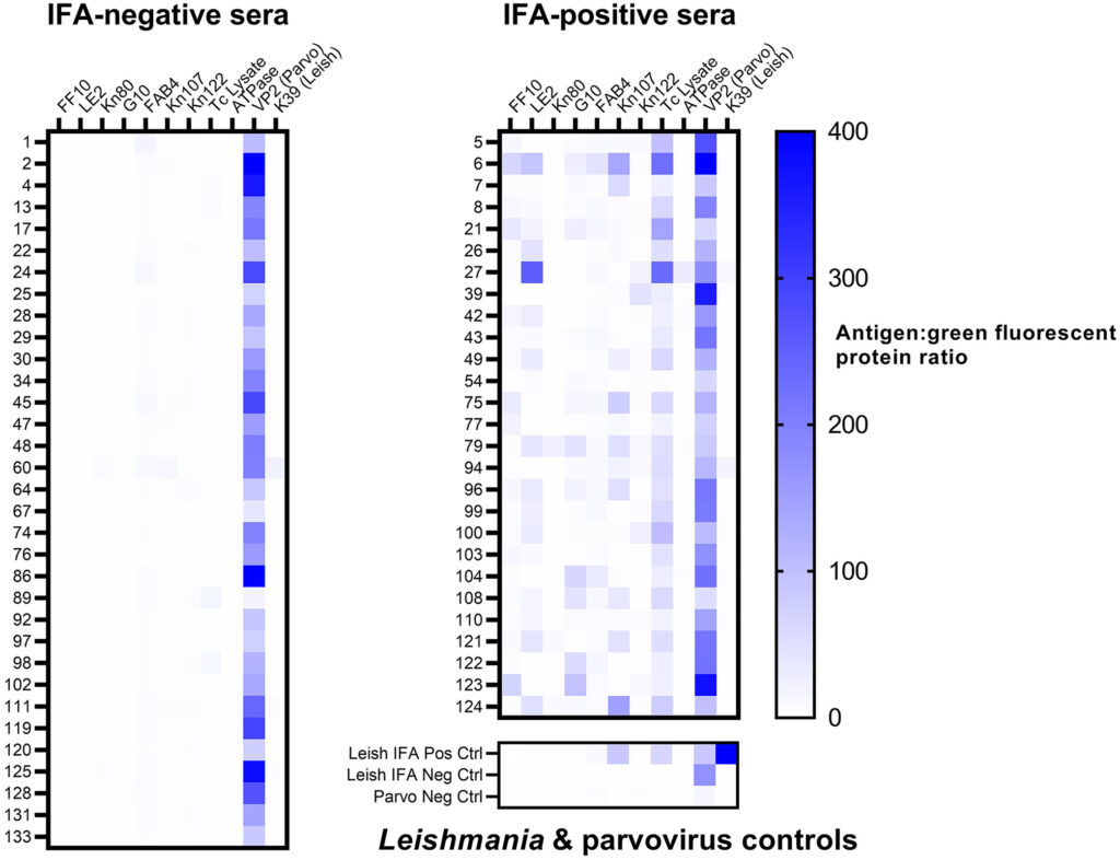
Figure 2. Heatmap of multiplex microsphere immunoassay reactivity of 60 canine serum previously tested for Trypanosoma cruzi antibodies by an indirect fluorescent antibody (IFA) test (grouped by previous IFA result), and reactivity of Leishmania and canine parvovirus controls.

The vector-borne protozoan parasite Trypanosoma cruzi causes Chagas disease in humans, dogs, and many other mammalian hosts. Canine Chagas disease is increasingly diagnosed in dogs of the southern United States where triatomine insect vectors occur, and there are limited veterinary testing options; only the indirect fluorescent antibody (IFA) test is offered at a single accredited diagnostic laboratory. We evaluated a multiplex microsphere immunoassay (MIA) for the detection of antibodies against T. cruzi in dogs and compared it with existing serologic methods to establish cutoff values and relative sensitivity and specificity. We tested 135 canine sera that had been characterized using the IFA and off-label use of 2 commercial rapid assays with our multiplex MIA against 12 antigens: 9 T. cruzi antigens, a negative control recombinant protein (green fluorescent protein, GFP), a Leishmania antigen, and a canine parvovirus antigen (used as an antibody control given near-ubiquitous parvoviral vaccination). The median fluorescence intensity (MFI) ratio between each T. cruzi antigen and GFP was calculated for every sample. Samples with an antigen:GFP MFI ratio > 4 SDs above the mean of 25 known-negative sera were considered positive to that antigen. Samples testing positive to ≥ 2 antigens were considered positive for T. cruzi antibodies. Compared to the IFA, our multiplex MIA had a relative sensitivity of 100% and specificity of 97.0%. Given its precision, high-throughput format, potential for automation, and lack of subjective interpretation, our multiplex MIA should be considered a valid and improved assay for T. cruzi antibodies in dogs.

Carlos A Rodriguez, Rachel E Busselman, Huifeng Shen, Ashley B Saunders, Rick Tarleton, Sarah A Hamer. J Vet Diagn Invest. 2023 Sep 5;10406387231198525. doi: 10.1177/10406387231198525.