MICU1 and MICU2 Play an Essential Role in Mitochondrial Ca2+ Uptake, Growth, and Infectivity of the Human Pathogen Trypanosoma cruzi
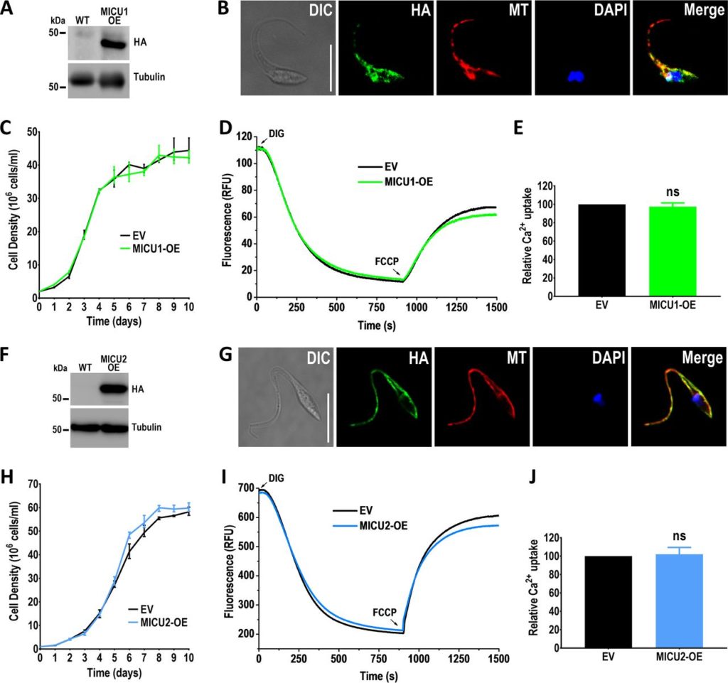 The mitochondrial Ca2+ uptake in trypanosomatids, which belong to the eukaryotic supergroup Excavata, shares biochemical characteristics with that of animals, which, together with fungi, belong to the supergroup Opisthokonta. However, the composition of the mitochondrial calcium uniporter (MCU) complex in trypanosomatids is quite peculiar, suggesting lineage-specific adaptations. In this work, we used Trypanosoma cruzi to study the role of orthologs for mitochondrial calcium uptake 1 (MICU1) and MICU2 in mitochondrial Ca2+ uptake. T. cruzi MICU1 (TcMICU1) and TcMICU2 have mitochondrial targeting signals, two canonical EF-hand calcium-binding domains, and localize to the mitochondria. Using the CRISPR/Cas9 system (i.e., clustered regularly interspaced short palindromic repeats with Cas9), we generated TcMICU1 and TcMICU2 knockout (-KO) cell lines. Ablation of either TcMICU1 or TcMICU2 showed a significantly reduced mitochondrial Ca2+uptake in permeabilized epimastigotes without dissipation of the mitochondrial membrane potential or effects on the AMP/ATP ratio or citrate synthase activity. However, none of these proteins had a gatekeeper function at low cytosolic Ca2+ concentrations ([Ca2+]cyt), as occurs with their mammalian orthologs. TcMICU1-KO and TcMICU2-KO epimastigotes had a lower growth rate and impaired oxidative metabolism, while infective trypomastigotes have a reduced capacity to invade host cells and to replicate within them as amastigotes. The findings of this work, which is the first to study the role of MICU1 and MICU2 in organisms evolutionarily distant from animals, suggest that, although these components were probably present in the last eukaryotic common ancestor (LECA), they developed different roles during evolution of different eukaryotic supergroups. The work also provides new insights into the adaptations of trypanosomatids to their particular life styles.
IMPORTANCE Trypanosoma cruzi is the etiologic agent of Chagas disease and belongs to the early-branching eukaryotic supergroup Excavata. Its mitochondrial calcium uniporter (MCU) subunit shares similarity with the animal ortholog that was important to discover its encoding gene. In animal cells, the MICU1 and MICU2 proteins act as Ca2+ sensors and gatekeepers of the MCU, preventing Ca2+ uptake under resting conditions and favoring it at high cytosolic Ca2+ concentrations ([Ca2+]cyt). Using the CRISPR/Cas9 technique, we generated TcMICU1 and TcMICU2 knockout cell lines and showed that MICU1 and -2 do not act as gatekeepers at low [Ca2+]cyt but are essential for normal growth, host cell invasion, and intracellular replication, revealing lineage-specific adaptations.
Mayara S. Bertolini, Miguel A. Chiurillo, Noelia Lander, Anibal E. Vercesi, Roberto Docampo. 2019. MBio.; 10(3). pii: e00348-19. doi: 10.1128/mBio.00348-19.
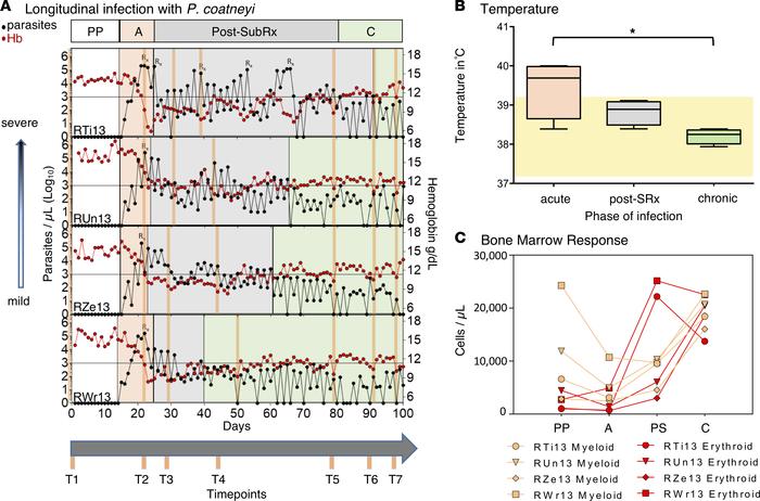 Chronic malaria is a major public health problem and significant challenge for disease eradication efforts. Despite its importance, the biological factors underpinning chronic malaria are not fully understood. Recent studies have shown that host metabolic state can influence malaria pathogenesis and transmission, but its role in chronicity is not known. Here, with the goal of identifying distinct modifications in the metabolite profiles of acute versus chronic malaria, metabolomics was performed on plasma from Plasmodium-infected humans and nonhuman primates with a range of parasitemias and clinical signs. In rhesus macaques infected with Plasmodium coatneyi, significant alterations in amines, carnitines, and lipids were detected during a high parasitemic acute phase and many of these reverted to baseline levels once a low parasitemic chronic phase was established. Plasmodium gene expression, studied in parallel in the macaques, revealed transcriptional changes in amine, fatty acid, lipid and energy metabolism genes, as well as variant antigen genes. Furthermore, a common set of amines, carnitines, and lipids distinguished acute from chronic malaria in plasma from human Plasmodium falciparum cases. In summary, distinct host-parasite metabolic environments have been uncovered that characterize acute versus chronic malaria, providing insights into the underlying host-parasite biology of malaria disease progression.
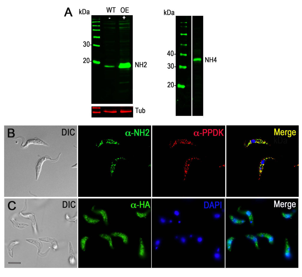 Trypanosoma brucei, a protist parasite that causes African trypanosomiasis or sleeping sickness, relies mainly on glycolysis for ATP production when in its mammalian host. Glycolysis occurs within a peroxisome-like organelle named the glycosome. Previous work from our laboratory reported the presence of significant amounts of inorganic polyphosphate (polyP), a polymer of three to hundreds of orthophosphate units, in the glycosomes and nucleoli of T. brucei In this work, we identified and characterized the activity of two Nudix hydrolases, TbNH2 and TbNH4, one located in the glycosomes and the other in the cytosol and nucleus, respectively, that can degrade polyP. We found that TbNH2 is an exopolyphosphatase with higher activity on short chain polyP, while TbNH4 is an endo- and exopolyphosphatase that has similar activity on polyP of various chain sizes. Both enzymes have higher activity at around pH 8.0. We also found that only TbNH2 can dephosphorylate ATP and ADP but with lower affinity than for polyP. Our results suggest that Nudix hydrolases can participate in polyP homeostasis and therefore may help control polyP levels in glycosomes, cytosol and nuclei of T. brucei.
 The macrocyclic lactone anthelmintics are the only class of drug currently used to prevent heartworm disease. Their extremely high potency in vivo is not mirrored by their activity against Dirofilaria immitis larvae in vitro, leading to suggestions that they may require host immune functions to kill the parasites. We have previously shown that ivermectin stimulates the binding of canine peripheral blood mononuclear cells (PBMCs) and polymorphonuclear leukocytes (PMNs) to D. immitis microfilariae (Mf). We have now extended these studies to moxidectin and examined the ability of both drugs to stimulate canine PBMC and PMN attachment to Mf from multiple strains of D. immitis, including two that are proven to be resistant to ivermectin in vivo. Both ivermectin and moxidectin significantly increased the percentage of drug-susceptible parasites with cells attached at very low concentrations (<10 nM), but much higher concentrations of ivermectin (>100 nM) were required to increase the percentage of the two resistant strains, Yazoo-2013 and Metairie-2014, with cells attached. Moxidectin increased the percentage of the two resistant strains with cells attached at lower concentrations (<10 nM) than did ivermectin. The attachment of the PBMCs and PMNs did not result in any parasite killing in vitro. These data support the biological relevance of the drug-stimulated attachment of canine leukocytes to D. immitis Mf and suggest that this phenomenon is related to the drug resistance status of the parasites.
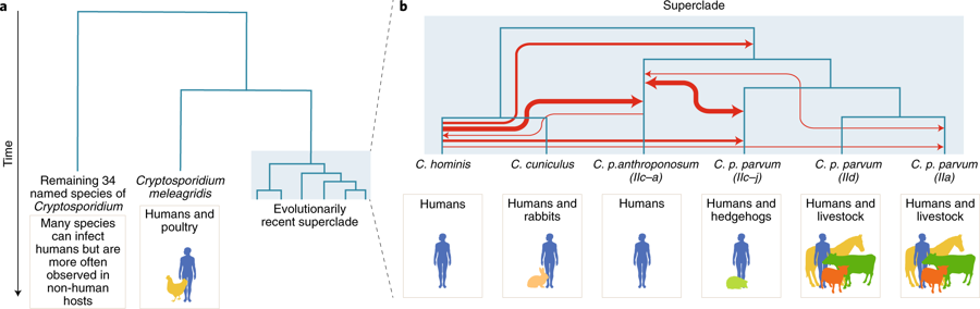 A large-scale comparative genomic survey of Cryptosporidium species and subtypes reveals a cryptic anthroponotic Cryptosporidium parvum branch and a large, recent superclade of species and subtypes that undergo genetic exchange, potentially facilitating host associations.
 Marc-Jan Gubbels received his Ph.D. at the University of Utrecht, the Netherlands, on diagnostics and vaccines for tick-transmitted Theileria and Babesia parasites of cattle. He then came to the CTEDG for a post-doc with Dr. Boris Striepen on Toxoplasma gondii. In 2005, he transitioned into an independent faculty position at Boston College and continued working on Toxoplasma. His lab is using and developing forward, reverse and functional genetic tools using enzymatic as well as fluorescent protein reporter assays in combination with cell sorting and fluorescence microscopy to learn more about the parasite’s cell biology.
 Macrophages are the major cell type infected by Leishmania parasites and the goal of my research is to define the relationship between Leishmania parasites, macrophages and the vasculature. Throughout my graduate studies and postdoctoral positions, I have dedicated my efforts toward understanding parasite-host interactions. As a graduate student at the University of Georgia carrying out research at the Centers for Disease Control and Prevention under Dr.
  James Morris is currently a Professor of Genetics and Biochemistry at Clemson University. He earned his BS at The College of William and Mary in Williamsburg VA in 1990 and an M.S. in Entomology at the University of Georgia in Athens GA in 1992. He completed his Ph.D. in Cellular Biology at the University of Georgia in 1997, characterizing the enzymology of a glycosylphosphatidylinositol phospholipase C from the African trypanosome, Trypanosoma brucei, under the supervision of Dr.
  Matthew Collins obtained his Ph.D. studying under mentor Dr.