Generic selectors
Exact matches only
Search in title
Search in content
Post Type Selectors

Author: Donna Huber

Riboflavin instability is a key factor underlying the requirement of a gut microbiota for mosquito development

We previously determined that several diets used to rear Aedes aegypti and other mosquito species support the development of larvae with a gut microbiota but do not support the development of axenic larvae. In contrast, axenic larvae have been shown to develop when fed other diets. To understand the mechanisms underlying this dichotomy, we developed a defined diet that could be manipulated in concert with microbiota composition and environmental conditions. Initial studies showed that axenic larvae could not grow under standard rearing conditions (27 °C, 16-h light: 8-h dark photoperiod) when fed a defined diet but could develop when maintained in darkness. Downstream assays identified riboflavin decay to lumichrome as the key factor that prevented axenic larvae from growing under standard conditions, while gut community members like Escherichia coli rescued development by being able to synthesize riboflavin. Earlier results showed that conventional and gnotobiotic but not axenic larvae exhibit midgut hypoxia under standard rearing conditions, which correlated with activation of several pathways with essential growth functions. In this study, axenic larvae in darkness also exhibited midgut hypoxia and activation of growth signaling but rapidly shifted to midgut normoxia and arrested growth in light, which indicated that gut hypoxia was not due to aerobic respiration by the gut microbiota but did depend on riboflavin that only resident microbes could provide under standard conditions. Overall, our results identify riboflavin provisioning as an essential function for the gut microbiota under most conditions A. aegypti larvae experience in the laboratory and field.

Yin Wang, Jai Hoon Eum, Ruby E. Harrison, Luca Valzania, Xiushuai Yang, Jena A. Johnson, Derek T. Huck, Mark R. Brown, Michael R. Strand Proceedings of the National Academy of Sciences Apr 2021, 118 (15) e2101080118; DOI: 10.1073/pnas.2101080118

Estimating true prevalence of Schistosoma mansoni from population summary measures based on the Kato-Katz diagnostic technique

Background: The prevalence of Schistosoma mansoni infection is usually assessed by the Kato-Katz diagnostic technique. However, Kato-Katz thick smears have low sensitivity, especially for light infections. Egg count models fitted on individual level data can adjust for the infection intensity-dependent sensitivity and estimate the ‘true’ prevalence in a population. However, application of these models is complex and there is need for adjustments that can be done without modelling expertise. This study provides estimates of the ‘true’ S. mansoni prevalence from population summary measures of observed prevalence and infection intensity using extensive simulations parametrized with data from different settings in sub-Saharan Africa.

Methodology: An individual-level egg count model was applied to Kato-Katz data to determine the S. mansoni infection intensity-dependent sensitivity for various sampling schemes. Observations in populations with varying forces of transmission were simulated, using standard assumptions about the distribution of worms and their mating behavior. Summary measures such as the geometric mean infection, arithmetic mean infection, and the observed prevalence of the simulations were calculated, and parametric statistical models fitted to the summary measures for each sampling scheme. For validation, the simulation-based estimates are compared with an observational dataset not used to inform the simulation.

Principal findings: Overall, the sensitivity of Kato-Katz in a population varies according to the mean infection intensity. Using a parametric model, which takes into account different sampling schemes varying from single Kato-Katy to triplicate slides over three days, both geometric and arithmetic mean infection intensities improve estimation of sensitivity. The relation between observed and ‘true’ prevalence is remarkably linear and triplicate slides per day on three consecutive days ensure close to perfect sensitivity.

Conclusions/significance: Estimation of ‘true’ S. mansoni prevalence is improved when taking into account geometric or arithmetic mean infection intensity in a population. We supply parametric functions and corresponding estimates of their parameters to calculate the ‘true’ prevalence for sampling schemes up to 3 days with triplicate Kato-Katz thick smears per day that allow estimation of the ‘true’ prevalence.

Bärenbold O, Garba A, Colley DG, Fleming FM, Assaré RK, Tukahebwa EM, et al. (2021) Estimating true prevalence of Schistosoma mansoni from population summary measures based on the Kato-Katz diagnostic technique. PLoS Negl Trop Dis 15(4): e0009310. https://doi.org/10.1371/journal.pntd.0009310

Mitochondrial Pyruvate Carrier Subunits Are Essential for Pyruvate-Driven Respiration, Infectivity, and Intracellular Replication of Trypanosoma cruzi

Pyruvate is the final metabolite of glycolysis and can be converted into acetyl coenzyme A (acetyl-CoA) in mitochondria, where it is used as the substrate for the tricarboxylic acid cycle. Pyruvate availability in mitochondria depends on its active transport through the heterocomplex formed by the mitochondrial pyruvate carriers 1 and 2 (MPC1/MPC2). We report here studies on MPC1/MPC2 of Trypanosoma cruzi, the etiologic agent of Chagas disease. Endogenous tagging of T. cruzi MPC1 (TcMPC1) and TcMPC2 with 3×c-Myc showed that both encoded proteins colocalize with MitoTracker to the mitochondria of epimastigotes. Individual knockout (KO) of TcMPC1 and TcMPC2 genes using CRISPR/Cas9 was confirmed by PCR and Southern blot analyses. Digitonin-permeabilized TcMPC1-KO and TcMPC2-KO epimastigotes showed reduced O2 consumption rates when pyruvate, but not succinate, was used as the mitochondrial substrate, while α-ketoglutarate increased their O2 consumption rates due to an increase in α-ketoglutarate dehydrogenase activity. Defective mitochondrial pyruvate import resulted in decreased Ca2+ uptake. The inhibitors UK5099 and malonate impaired pyruvate-driven oxygen consumption in permeabilized control cells. Inhibition of succinate dehydrogenase by malonate indicated that pyruvate needs to be converted into succinate to increase respiration. TcMPC1-KO and TcMPC2-KO epimastigotes showed little growth differences in standard or low-glucose culture medium. However, the ability of trypomastigotes to infect tissue culture cells and replicate as intracellular amastigotes was decreased in TcMPC-KOs. Overall, T. cruzi MPC1 and MPC2 are essential for cellular respiration in the presence of pyruvate, invasion of host cells, and replication of amastigotes.

IMPORTANCE Trypanosoma cruzi is the causative agent of Chagas disease. Pyruvate is the end product of glycolysis, and its transport into the mitochondrion is mediated by the mitochondrial pyruvate carrier (MPC) subunits. Using the CRISPR/Cas9 technique, we generated individual T. cruzi MPC1 (TcMPC1) and TcMPC2 knockouts and demonstrated that they are essential for pyruvate-driven respiration. Interestingly, although glycolysis was reported as not an important source of energy for the infective stages, MPC was essential for normal host cell invasion and intracellular replication.

Raquel S. NegreirosNoelia LanderMiguel A. ChiurilloAnibal E. VercesiRoberto Docampo.

Synthesis of Mono- and Bisperoxide-Bridged Artemisinin Dimers to Elucidate the Contribution of Dimerization to Antimalarial Activity

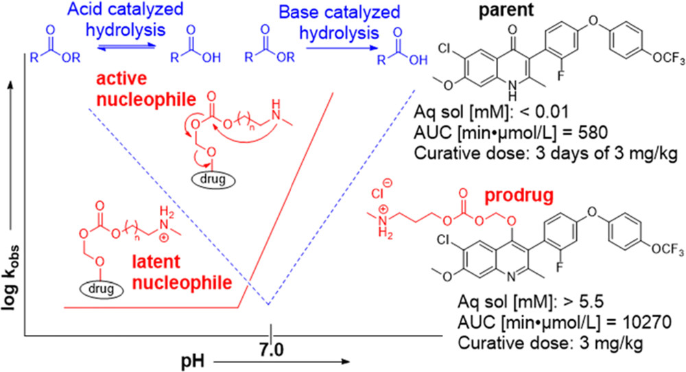
During the past decade, artemisinin as an antimalarial has been in the spotlight, in part due to the Nobel Prize in Physiology or Medicine awarded to Tu Youyou. While many studies have been completed detailing the significant increase in activity resulting from the dimerization of natural product artemisinin, activity increases unaccounted for by the peroxide bridge have yet to be researched. Here we outline the synthesis and testing for antimalarial activity of artemisinin dimers in which the peroxide bridge in one-half of the dimer is reduced, resulting in a dimer with one active and one deactivated artemisinin moiety.

Cynthia L Lichorowic, Yingzhao Zhao, Steven P Maher, Vivian Padín-Irizarry, Victoria C Mendiola, Sagan T de Castro, Jacob A Worden, Debora Casandra, Dennis E Kyle, Roman Manetsch. ACS Infect Dis. 2021 Apr 1. doi: 10.1021/acsinfecdis.1c00066

Ca2+ entry at the plasma membrane and uptake by acidic stores is regulated by the activity of the V‐H+‐ATPase in Toxoplasma gondii

Ca2+ is a universal intracellular signal that regulates many cellular functions. In Toxoplasma gondii, the controlled influx of extracellular and intracellular Ca2+ into the cytosol initiates a signaling cascade that promotes pathogenic processes like tissue destruction and dissemination. In this work we studied the role of proton transport in cytosolic Ca2+ homeostasis and the initiation of Ca2+ signaling. We used a T. gondii mutant of the V-ATPase, a pump previously shown to transport protons to the extracellular medium, control intracellular pH and membrane potential and we show that proton gradients are important for maintaining resting cytosolic Ca2+ at physiological levels and for Ca2+ influx. Proton transport was also important for Ca2+ storage by acidic stores and, unexpectedly, the endoplasmic reticulum. Proton transport impacted the amount of polyphosphate (polyP), a phosphate polymer that binds Ca2+ and concentrate in acidocalcisomes. This was supported by the co-localization of the vacuolar transporter chaperone 4 (VTC4), the catalytic subunit of the VTC complex that synthesizes polyP, with the V-ATPase in acidocalcisomes. Our work show that proton transport regulate plasma membrane Ca2+ transport and control acidocalcisome polyP and Ca2+ content impacting Ca2+ signaling and downstream stimulation of motility and egress in T. gondii.

Andrew J Stasic, Eric J Dykes, Ciro D Cordeiro, Stephen A Vella, Mojtaba S Fazli, Shannon Quinn, Roberto Docampo, Silvia N J Moreno. Mol Microbiol. 2021 Apr 1. doi: 10.1111/mmi.14722

High variation in immune responses and parasite phenotypes in naturally acquired Trypanosoma cruzi infection in a captive non-human primate breeding colony in Texas, USA

Trypanosoma cruzi, the causative agent of human Chagas disease, is endemic to the southern region of the United States where it routinely infects many host species. The indoor/outdoor housing configuration used in many non-human primate research and breeding facilities in the southern of the USA provides the opportunity for infection by T. cruzi and thus provides source material for in-depth investigation of host and parasite dynamics in a natural host species under highly controlled and restricted conditions. For cynomolgus macaques housed at such a facility, we used a combination of serial blood quantitative PCR (qPCR) and hemoculture to confirm infection in >92% of seropositive animals, although each method alone failed to detect infection in >20% of cases. Parasite isolates obtained from 43 of the 64 seropositive macaques were of 2 broad genetic types (discrete typing units, (DTU’s) I and IV); both within and between these DTU groupings, isolates displayed a wide variation in growth characteristics and virulence, elicited host immune responses, and susceptibility to drug treatment in a mouse model. Likewise, the macaques displayed a diversity in T cell and antibody response profiles that rarely correlated with parasite DTU type, minimum length of infection, or age of the primate. This study reveals the complexity of infection dynamics, parasite phenotypes, and immune response patterns that can occur in a primate group, despite being housed in a uniform environment at a single location, and the limited time period over which the T. cruzi infections were established.

Padilla AM, Yao PY, Landry TJ, Cooley GM, Mahaney SM, Ribeiro I, VandeBerg JL. Tarleton RL. (2021) High variation in immune responses and parasite phenotypes in naturally acquired Trypanosoma cruzi infection in a captive non-human primate breeding colony in Texas, USA. PLoS Negl Trop Dis 15(3): e0009141. https://doi.org/10.1371/journal.pntd.0009141

Naegleria fowleri: Protein structures to facilitate drug discovery for the deadly, pathogenic free-living amoeba

Naegleria fowleri is a pathogenic, thermophilic, free-living amoeba which causes primary amebic meningoencephalitis (PAM). Penetrating the olfactory mucosa, the brain-eating amoeba travels along the olfactory nerves, burrowing through the cribriform plate to its destination: the brain’s frontal lobes. The amoeba thrives in warm, freshwater environments, with peak infection rates in the summer months and has a mortality rate of approximately 97%. A major contributor to the pathogen’s high mortality is the lack of sensitivity of N. fowleri to current drug therapies, even in the face of combination-drug therapy. To enable rational drug discovery and design efforts we have pursued protein production and crystallography-based structure determination efforts for likely drug targets from N. fowleri. The genes were selected if they had homology to drug targets listed in Drug Bank or were nominated by primary investigators engaged in N. fowleri research. In 2017, 178 N. fowleri protein targets were queued to the Seattle Structural Genomics Center of Infectious Disease (SSGCID) pipeline, and to date 89 soluble recombinant proteins and 19 unique target structures have been produced. Many of the new protein structures are potential drug targets and contain structural differences compared to their human homologs, which could allow for the development of pathogen-specific inhibitors. Five of the structures were analyzed in more detail, and four of five show promise that selective inhibitors of the active site could be found. The 19 solved crystal structures build a foundation for future work in combating this devastating disease by encouraging further investigation to stimulate drug discovery for this neglected pathogen.

Logan Tillery, Kayleigh Barrett, Jenna Goldstein, Jared W Lassner, Bram Osterhout, Nathan L Tran, Lily Xu, Ryan M Young, Justin Craig, Ian Chun, David M Dranow, Jan Abendroth, Silvia L Delker, Douglas R Davies, Stephen J Mayclin, Brandy Calhoun, Madison J Bolejack, Bart Staker, Sandhya Subramanian, Isabelle Phan, Donald D Lorimer, Peter J Myler, Thomas E Edwards, Dennis E Kyle, Christopher A Rice, James C Morris, James W Leahy, Roman Manetsch, Lynn K Barrett, Craig L Smith, Wesley C Van Voorhis (2021) Naegleria fowleri: Protein structures to facilitate drug discovery for the deadly, pathogenic free-living amoeba. PLoS ONE 16(3): e0241738. https://doi.org/10.1371/journal.pone.0241738

UGA’s Rozario receives NIH Director’s New Innovator Award

By Alan Flurry

(Photo courtesy of Tania Rozario)

University of Georgia faculty member Tania Rozario has received a $2 million grant from the National Institutes of Health Director’s New Innovator Award Program, which supports early-career investigators of exceptional creativity who propose high-risk, high-reward research projects.

Rozario is an assistant professor with a joint appointment in the Franklin College of Arts and Sciences Department of Genetics and the Center for Tropical and Emerging Global Diseases.

Among the study of tropical diseases worldwide—and particularly among the parasites that cause disease—worms are a largely neglected disease agent, despite being a source of widespread problems that affect both health and economic output. Even within the study of worms, parasitic flatworms like tapeworms represent an understudied group. However, free-living flatworms like planarians are the focus of significant research because of the organism’s dynamic regenerative capacity, which presents intriguing parallels to their parasitic cousins.

Planarian flatworms cut in two will make two new worms, and cut into 10 pieces will result in 10 worms. They are the Ferrari of regenerators, according to Rozario.

“As part of its normal life cycle, a tapeworm sheds large parts of its body and then regrows this lost tissue,” Rozario said. “It has this natural regenerative-like ability, which is very promising from a basic biology standpoint, to understand how stem cells and regeneration functions in these worms.”

Taking advantage of both extensive past research and the much more sophisticated tools of today, Rozario envisions a melding of developmental biology with parasitology as a new approach to understand the parasite. She is using the rat tapeworm, Hymenolepis diminuta, to re-establish a model organism that had been a favorite model among parasitologists in the early-mid 20th century but was left behind by the molecular biology revolution.

Flatworms have incredible capacity for regeneration, according to Tania Rozario, who studies them as agents of parasitic disease. For example, this rat tapeworm is capable of growing thousands of segments and can regenerate segments following amputation.


“One of the major drivers is trying to understand the diversity of the stem cell milieu in these tapeworms,” Rozario said. “We’ll try to parse out the interaction between the diversity of stem cells present and the local signals that then allow the worms to regenerate and make thousands of segments. These interactions are likely crucial for development of both female and male reproductive structures, which exist in each segment of the animal.”

“Dr. Rozario brings a new and exciting area of research to UGA, and her enthusiasm for her research is phenomenal,” said Nancy Manley, Distinguished Research Professor and department head for genetics. “Her success in getting this prestigious award speaks to her talent and the quality of her science. We are enthusiastic to have her as our newest colleague.”

“I am excited that Dr. Rozario has joined us at UGA,” said Dennis Kyle, GRA Eminent Scholar in antiparasitic drug discovery and director of the Center for Tropical and Emerging Global Diseases. “Her pioneering work is ushering in a new era whereby tapeworms can serve as model organisms. The prestigious NIH Director’s Pioneer Award is evidence of the creative approach she is undertaking to better understand these interesting parasites.”

“It’s important that we study the monsters in our midst so that we can learn from organisms in our environment that have these really out-there, unique physiological capabilities,” Rozario said. “We can learn about how they have evolved strategies to thrive in their specific niche, but they can also teach us something more fundamental about biology that could be broadly applicable.”

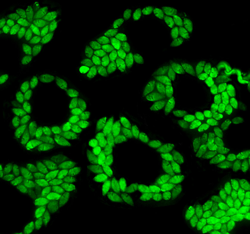
microscopy of tapeworms
In these Hymenolepis diminuta tapeworm necks, dividing cells—including stem cells—are depicted through color-marking in the image. (Image courtesy of Tania Rozario)

Lysosomal cholesterol accumulation contributes to the movement phenotypes associated with NUS1 haploinsufficiency

Purpose: Variants in NUS1 are associated with a congenital disorder of glycosylation, developmental and epileptic encephalopathies, and are possible contributors to Parkinson disease pathogenesis. How the diverse functions of the NUS1-encoded Nogo B receptor (NgBR) relate to these different phenotypes is largely unknown. We present three patients with de novo heterozygous variants in NUS1 that cause a complex movement disorder, define pathogenic mechanisms in cells and zebrafish, and identify possible therapy.

Methods: Comprehensive functional studies were performed using patient fibroblasts, and a zebrafish model mimicking NUS1 haploinsufficiency.

Results: We show that de novo NUS1 variants reduce NgBR and Niemann-Pick type C2 (NPC2) protein amount, impair dolichol biosynthesis, and cause lysosomal cholesterol accumulation. Reducing nus1 expression 50% in zebrafish embryos causes abnormal swim behaviors, cholesterol accumulation in the nervous system, and impaired turnover of lysosomal membrane proteins. Reduction of cholesterol buildup with 2-hydroxypropyl-ß-cyclodextrin significantly alleviates lysosomal proteolysis and motility defects.

Conclusion: Our results demonstrate that these NUS1 variants cause multiple lysosomal phenotypes in cells. We show that the movement deficits associated with nus1 reduction in zebrafish arise in part from defective efflux of cholesterol from lysosomes, suggesting that treatments targeting cholesterol accumulation could be therapeutic.

Seok-Ho Yu, Tong Wang, Kali Wiggins, Raymond J. Louie, Emilio F. Merino, Cindy Skinner, Maria B. Cassera, Kirsten Meagher, Paul Goldberg, Neggy Rismanchi, Dillon Chen, Michael J. Lyons, Heather Flanagan-Steet & Richard Steet. Genet Med. 2021 Mar 17. doi: 10.1038/s41436-021-01137-6.