Significance: Millions of people are infected with trypanosomatids and new therapeutic approaches are needed. Trypanosomatids possess one mitochondrion per cell, and its study has led to discoveries of general biological interest. These mitochondria, as their animal counterparts, generate reactive oxygen species (ROS) and have enzymatic and non-enzymatic defenses against them. Mitochondrial calcium ion (Ca2+) overload leads to the generation of ROS and its study could lead to relevant information on the biology of trypanosomatids and to novel drug targets. Recent Advances: Mitochondrial Ca2+ is normally involved in maintaining the bioenergetics of trypanosomes but when Ca2+ overload occurs it is associated to cell death. Trypanosomes lack key players of the mechanism of cell death described in mammalian cells although mitochondrial Ca2+ overload results in collapse of their membrane potential, production of ROS, and cytochrome c release. They are also very resistant to mitochondrial permeability transition, and cell death after mitochondrial Ca2+ overload depends on the generation of ROS.
Critical issues: In this review, we consider the mechanisms of mitochondrial oxidant generation and removal, and the involvement of Ca2+ in trypanosome cell death.
Future directions: More studies are required to determine the reactions involved in the generation of ROS by the mitochondria of trypanosomatids, their enzymatic and non-enzymatic defenses against ROS, and the occurrence and composition of a mitochondrial permeability transition pore.
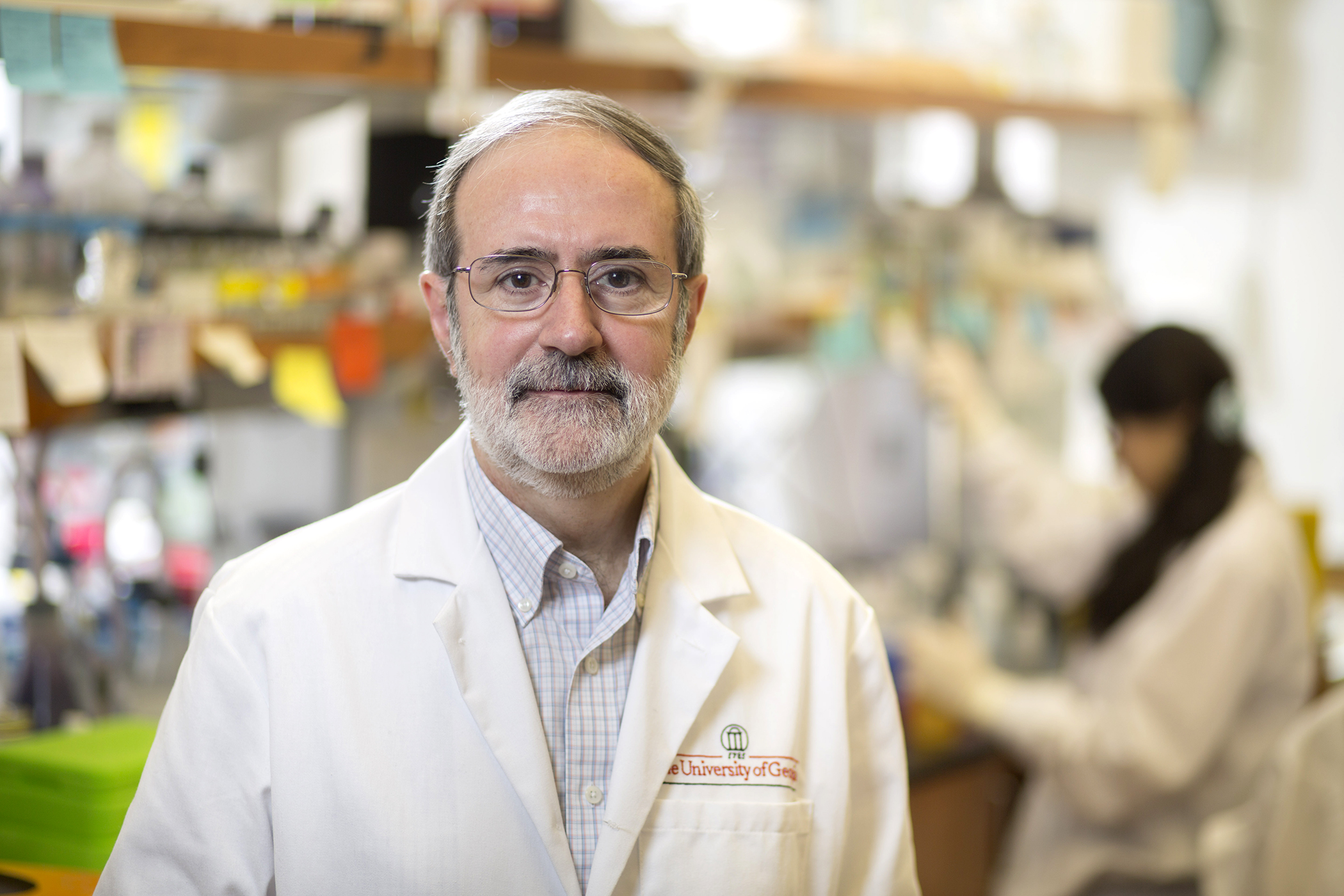
Ronald Drew Etheridge’s scientific career can be characterized by one word—serendipity.
After completing his bachelor’s degree in biochemistry and molecular biology with a Spanish language minor at the University of Georgia, Etheridge set out for Spain, where he traveled and worked as an English teacher. On his return home, and in need of a job, a former coworker mentioned a potential opening for a technician at UGA in the lab of Rick Tarleton, a leader in studying Trypanosoma cruzi, the protozoan that causes Chagas’ disease. While having worked in many labs as an undergraduate conducting basic scientific research, he had never really considered pursuing a study of immunology or parasitology. As luck would have it, his time in the Tarleton lab would spark his scientific curiosity like never before.
“It was the first time science was truly fun for me,” said Etheridge. “I really enjoyed the interesting scientific debates and rigorous research environment fostered in Rick’s lab.”
Realizing he needed further training to be a competent parasitologist, he went on to pursue a Ph.D. at the University of California, Irvine, and postdoctoral training at Washington University School of Medicine. In 2016, Etheridge returned to his alma mater and joined the faculty in Franklin College of Arts and Science’s Department of Cellular Biology and the Center for Tropical and Emerging Global Disease as an assistant professor.
By the time he returned to UGA, his focus had shifted slightly from immunology to molecular parasitology as he delved into host-pathogen interactions involving the protozoan parasite Toxoplasma gondii. But serendipity struck again. Upon his return to UGA he realized that Tarleton and colleague Roberto Docampo had pioneered the use of the gene-editing system CRISPR-Cas9 in Trypanosoma cruzi. Their research opened up the possibility of studying this highly neglected parasite at the molecular level for the first time. This work ultimately led Etheridge to pilot gene-editing projects in T. cruzi with a focus on explaining how this parasite directly interacts with and manipulates its host.
“One of the great things about academic research is the ability to be flexible and go down new avenues of research when they present themselves,” said Etheridge.
As part of these pilot studies, Etheridge’s group identified the first protein components of what can be considered the digestive tract of this single-cell parasite. This unique feeding structure starts as a pore on the parasite surface (the cytostome) and is followed by a tubular structure called the cytopharynx that ultimately ends with captured food being sent for digestion in endocytosed vesicles. The Etheridge lab refers to this endocytic feeding organelle as the cytostome/cytopharynx complex, or SPC for short.
“That’s what is cool about science—by chance you find novel things,” said Etheridge.
When this project began, very little was known about how T. cruzi fed on its host to obtain nutrients. Since this initial discovery, the Etheridge lab has identified dozens of SPC-targeted proteins and has uncovered the protein machinery parasites use to catch and bring in food they want to digest.
“Virtually nothing was known about how this structure actually worked,” said Etheridge. “There have been some electron microscopy studies that described the structure, but that’s all we had when we first started. It has been really exciting to work on something so fundamental yet so poorly understood.”
The National Institutes of Health awarded Etheridge a new five-year grant to continue down this path in hopes of deciphering how the SPC works and the role this structure plays in T. cruzi’s parasitic life cycle. The answers to these questions could have wide implications.
“Not only can it help us to devise potential drug treatments for Chagas’ disease, an often debilitating and sometimes fatal disease which adversely affects 10 million people in the Americas,” said Etheridge. “But more broadly, it can also tell us something fundamental about the basic biology of many species of protozoa that also use the SPC structure to capture and digest food.”
Mayara Bertolini is a third year Ph.D. trainee in the laboratory of Dr. Roberto Docampo. She has recently been awarded a predoctoral fellowship from the American Heart Association.
Please tell us a little about yourself.
I am from São Paulo, Brazil and I have always been a very curious person that likes to discover unique things. Over time, I realized that biology was one of my favorite subjects, especially when it came to diseases. I decided to major in Biomedical Sciences at the Faculdade Anhanguera de Santa Bárbara D’Oeste (São Paulo, Brazil). After my graduation, I performed voluntary research training at the Laboratory of Bioenergetics of the Department of Clinical Pathology (School of Medical Sciences) of the State University of Campinas (Campinas, São Paulo, Brazil) under the supervision of Dr. Anibal Vercesi. Thereafter, I joined the Master’s program to continue my training as a scientist. There I met Dr. Roberto Docampo, who has collaborated with Dr. Vercesi for many years. Since then, I joined his research group, where Dr. Miguel Angel Chiurillo and Dr. Noelia Lander were also members of a very productive team, which has stimulated my fascination for research in parasitology. During my master’s, I was awarded a fellowship from the São Paulo Research Foundation (FAPESP) to perform a functional study of the regulatory subunits Mitochondrial Ca2+ Uptake 1 (MICU1) and 2 (MICU2) involved in calcium signaling in the parasite that causes Chagas disease, Trypanosoma cruzi. My master’s project elucidated some questions and opened doors to interesting new topics, which our group is very excited to explain.
Why did you choose UGA?
I wanted to continue working with the same model to improve my scientific thinking and to complete my laboratory training, and the Center for Tropical and Emerging Global Diseases (CTEGD) at UGA has a wide range of researchers working with trypanosomes. Pursuing my Ph.D. at UGA is an extraordinary opportunity because of CTEGD’s unique infrastructure, which consists of extremely qualified professionals and resources that facilitate the development of research projects.
What is your research focus?
T. cruzi is one of the least well understood neglected tropical disease agents and current treatments remain inadequate partly due to a general lack of knowledge of this parasite’s basic biology. We are particularly interested in establishing the role and interaction between mitochondrial proteins involved in Ca2+ uptake in this organelle. Understanding the mechanisms of adaptation and survival of the parasite upon environmental challenges, as changes in concentration of free Ca2+, will lead to important insights into the biology of this parasite and the evolution of Ca2+ signaling in eukaryotic cells. Considering that disruption of Ca2+ homeostasis by toxic agents is related to the loss of cell viability, the identification of the possible differences in mitochondrial Ca2+ transport between these parasites and the host cells could be useful for the development of new chemotherapeutic agents against Chagas disease. The purpose of the AHA predoctoral fellowship is to enhance the training of students who intend to pursue careers as scientists aimed at improving global health and wellbeing, and I feel like I can contribute to this mission.
What are your future professional plans?
After my graduation from UGA, I hope to continue for a postdoctoral research position. In the future, I would like to establish a research group in Brazil using trypanosomatids as biological models for studying the structure and function of proteins.
Any advice for a student interested in this field?
Don’t be afraid to try new things and learn from it.
Support trainees like Mayara by giving today to the Center for Tropical & Emerging Global Diseases.
Naegleria fowleri is a pathogenic free-living amoeba that is commonly found in warm freshwater and can cause a rapidly fulminant disease known as primary amoebic meningoencephalitis (PAM). New drugs are urgently needed to treat PAM, as the fatality rate is >97%. Until recently, few advances have been made in the discovery of new drugs for N. fowleri, and one drawback is the lack of validated tools and methods to enhance drug discovery and diagnostics research. In this study, we aimed to validate alternative methods to assess cell proliferation that are commonly used for other cell types and develop a novel drug screening assay to evaluate drug efficacy on N. fowleri replication. EdU (5-ethynyl-2′-deoxyuridine) is a pyrimidine analog of thymidine that can be used as a quantitative endpoint for cell proliferation. EdU incorporation is detected via a copper catalyzed click reaction with an Alexa Fluor-linked azide. EdU incorporation in replicating N. fowleri was validated using fluorescence microscopy, and quantitative methods for assessing EdU incorporation were developed by using an imaging flow cytometer. Currently used PAM therapeutics inhibited N. fowleri replication and EdU incorporation in vitro. EdA (7-deaza-2′-deoxy-7-ethynyladenosine), an adenine analog, also was incorporated by N. fowleri but was more cytotoxic than EdU. In summary, EdU incorporation could be used as a complimentary method for drug discovery for these neglected pathogens.
Emma V Troth, Dennis E Kyle. Antimicrob Agents Chemother. 2021 Jun 17;65(7):e0001721. doi: 10.1128/AAC.00017-21.
Plasmodium is a genus of apicomplexan parasites which replicate in the liver before causing malaria. Plasmodium vivax can also persist in the liver as dormant hypnozoites and cause clinical relapse upon activation, but the molecular mechanisms leading to activation have yet to be discovered. In this study, we use high-resolution microscopy to characterize temporal changes of the P. vivax liver stage tubovesicular network (TVN), a parasitophorous vacuole membrane (PVM)-derived network within the host cytosol. We observe extended membrane clusters, tubules, and TVN-derived vesicles present throughout P. vivax liver stage development. Additionally, we demonstrate an unexpected presence of the TVN in hypnozoites and observe some association of this network to host nuclei. We also reveal that the host water and solute channel aquaporin-3 (AQP3) associates with TVN-derived vesicles and extended membrane clusters. AQP3 has been previously shown to localize to the PVM of P. vivax hypnozoites and liver schizonts but has not yet been shown in association to the TVN. Our results highlight host-parasite interactions occur in both dormant and replicating liver stage P. vivax forms and implicate AQP3 function during this time. Together, these findings enhance our understanding of P. vivax liver stage biology through characterization of the TVN with an emphasis on the presence of this network in dormant hypnozoites.
Kayla Sylvester, Steven P Maher, Dora Posfai, Michael K Tran, McKenna C Crawford, Amélie Vantaux, Benoît Witkowski, Dennis E Kyle, Emily R Derbyshire. Front Cell Infect Microbiol. 2021 Jun 14;11:687019. doi: 10.3389/fcimb.2021.687019. eCollection 2021
Cryptosporidiosis is ranked sixth in the list of the most important food-borne parasites globally, and it is an important contributor to mortality in infants and the immunosuppressed. Recently, the number of genome sequences available for this parasite has increased drastically. The majority of the sequences are derived from population studies of Cryptosporidium parvum and Cryptosporidium hominis, the most important species causing disease in humans. Work with this parasite is challenging since it lacks an optimal, prolonged, in vitro culture system, which accurately reproduces the in vivo life cycle. This obstacle makes the cloning of isolates nearly impossible. Thus, patient isolates that are sequenced represent a population or, at times, mixed infections. Oocysts, the lifecycle stage currently used for sequencing, must be considered a population even if the sequence is derived from single-cell sequencing of a single oocyst because each oocyst contains four haploid meiotic progeny (sporozoites). Additionally, the community does not yet have a set of universal markers for strain typing that are distributed across all chromosomes. These variables pose challenges for population studies and require careful analyses to avoid biased interpretation. This review presents an overview of existing population studies, challenges, and potential solutions to facilitate future population analyses.
Transient Receptor Potential (TRP) channels participate in calcium ion (Ca2+) influx and intracellular Ca2+ release. TRP channels have not been studied in Toxoplasma gondii or any other apicomplexan parasite. In this work we characterize TgGT1_310560, a protein predicted to possess a TRP domain (TgTRPPL-2) and determined its role in Ca2+ signaling in T. gondii, the causative agent of toxoplasmosis. TgTRPPL-2 localizes to the plasma membrane and the endoplasmic reticulum (ER) of T. gondii. The ΔTgTRPPL-2 mutant was defective in growth and cytosolic Ca2+ influx from both extracellular and intracellular sources. Heterologous expression of TgTRPPL-2 in HEK-3KO cells allowed its functional characterization. Patching of ER-nuclear membranes demonstrates that TgTRPPL-2 is a non-selective cation channel that conducts Ca2+. Pharmacological blockers of TgTRPPL-2 inhibit Ca2+ influx and parasite growth. This is the first report of an apicomplexan ion channel that conducts Ca2+ and may initiate a Ca2+ signaling cascade that leads to the stimulation of motility, invasion and egress. TgTRPPL-2 is a potential target for combating Toxoplasmosis.
Karla Marie Marquez-Nogueras, Myriam Andrea Hortua Triana, Nathan M Chasen, Ivana Y Kuo, Silvia NJ Moreno. Elife. 2021 Jun 9;10:e63417. doi: 10.7554/eLife.63417.
Gut microbes and diet can both strongly affect the biology of multicellular animals, but it is often difficult to disentangle microbiota-diet interactions due to the complex microbial communities many animals harbor and the nutritionally variable diets they consume. While theoretical and empirical studies indicate that greater microbiota diversity is beneficial for many animal hosts, there have been few tests performed in aquatic invertebrates. Most mosquito species are aquatic detritivores during their juvenile stages that harbor variable microbiotas and consume diets that range from nutrient rich to nutrient poor. In this study, we produced a gnotobiotic model that allowed us to examine how interactions between specific gut microbes and diets affect the fitness of Aedes aegypti, the yellow fever mosquito. Using a simplified seven-member community of bacteria (ALL7) and various laboratory and natural mosquito diets, we allowed larval mosquitoes to develop under different microbial and dietary conditions and measured the resulting time to adulthood and adult size. Larvae inoculated with the ALL7 or a more complex community developed similarly when fed nutrient-rich rat chow or fish food laboratory diets, whereas larvae inoculated with individual bacterial members of the ALL7 community exhibited few differences in development when fed a rat chow diet but exhibited large differences in performance when fed a fish food diet. In contrast, the ALL7 community largely failed to support the growth of larvae fed field-collected detritus diets unless supplemented with additional protein or yeast. Collectively, our results indicate that mosquito development and fitness are strongly contingent on both diet and microbial community composition.
Dr. Dennis Kyle, director of CTEGD and professor in the departments of cellular biology and infectious diseases, is the featured guest on Episode 5 of the People, Parasites & Plagues Podcast. He talks about a deadly disease caused by Naegleria fowleri, also known as the brain-eating amoeba.
People, Parasites & Plagues is a podcast aimed at delivering information about the fascinating pathogens among us from the impressive professionals who study them.
Join hosts Dr. David Peterson and Dr. Liliana Salvador, two infectious disease researchers from the University of Georgia, as they explore the past, present, and future of science.
Tune in every other week for a new and enlightening episode as they unpack the details surrounding some of Earth’s most perplexing diseases. Look for the People, Parasites & Plagues Podcast on your favorite Podcast service!
Leucine zipper-EF-hand containing transmembrane protein 1 (Letm1) is a mitochondrial inner membrane protein involved in Ca2+ and K+ homeostasis in mammalian cells. Here, we demonstrate that the Letm1 orthologue of Trypanosoma cruzi, the etiologic agent of Chagas disease, is important for mitochondrial Ca2+ uptake and release. The results show that both mitochondrial Ca2+ influx and efflux are reduced in TcLetm1 knockdown (TcLetm1-KD) cells and increased in TcLetm1 overexpressing cells, without alterations in the mitochondrial membrane potential. Remarkably, TcLetm1 knockdown or overexpression increases or does not affect mitochondrial Ca2+ levels in epimastigotes, respectively. TcLetm1-KD epimastigotes have reduced growth, and both overexpression and knockdown of TcLetm1 cause a defect in metacyclogenesis. TcLetm1-KD also affected mitochondrial bioenergetics. Invasion of host cells by TcLetm1-KD trypomastigotes and their intracellular replication is greatly impaired. Taken together, our findings indicate that TcLetm1 is important for Ca2+ homeostasis and cell viability in T cruzi.
Guilherme Rodrigo Rm Dos Santos, Ana Catarina Rezende Leite, Noelia Lander, Miguel Angel Chiurillo, Aníbal Eugênio Vercesi, Roberto Docampo. FASEB J. 2021 Jul;35(7):e21685. doi: 10.1096/fj.202100120RR