Insulin-like peptides activate egg formation in the Asian malaria mosquito Anopheles stephensi
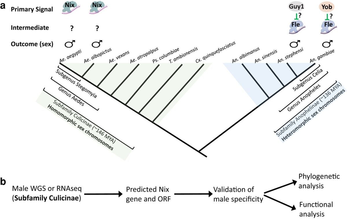
Background: The mosquito family Culicidae diverged into the subfamilies Anophelinae and Culicinae approximately 179 million years ago. Most female mosquitoes are anautogenous and must blood-feed on a vertebrate to produce eggs. Regulation of egg-producing gonotrophic cycles is best understood in the culicine Aedes aegypti. Anopheline mosquitoes encode all of the hormones that regulate gonotrophic cycles in …