Generic selectors
Exact matches only
Search in title
Search in content
Post Type Selectors

Tag: Don Harn

First bovine vaccine to prevent human schistosomiasis – a cluster randomised Phase 3 clinical trial

Objective

Schistosomiasis is a neglected tropical parasitic disease caused by blood flukes of the genus Schistosoma. Schistosoma japonicum is zoonotic in China, the Philippines, and Indonesia, with bovines acting as major reservoirs of human infection. The primary objective of the trial was to examine the impact of a combination of human mass chemotherapy, snail control through mollusciciding, and SjCTPI bovine vaccination on the rate of human infection.

Methods

A 5-year phase IIIa cluster randomized control trial was conducted among 18 schistosomiasis-endemic villages comprising 18,221 residents in Northern Samar, The Philippines.

Results

Overall, bovine vaccination resulted in a statistically significant decrease in human infection (relative risk [RR] = 0.75; 95% confidence interval [CI] = 0.69 to 0.82) across all trial follow-ups. The best outcome of the trial was when bovine vaccination was combined with snail mollusciciding. This combination resulted in a 31% reduction (RR = 0.69; 95% CI = 0.61 to 0.78) in human infection.

Conclusion

This is the first trial to demonstrate the effectiveness of a bovine vaccine for schistosomiasis in reducing human schistosome infection. The trial is registered with Australian New Zealand Clinical Trials Registry (ACTRN12619001048178).

Allen G. Ross, Donald A. Harn, Delia Chy, Marianette Inobaya, Jerric R. Guevarra, Lisa Shollenberger, Yuesheng Li, Donald P. McManus, Darren J. Gray and Gail M. Williams. 2023. International Journal of Infectious Diseases. S1201-9712(23)00038-3. doi: 10.1016/j.ijid.2023.01.037. Online ahead of print.

Evaluation of delayed LNFPIII treatment initiation protocol on improving long-term behavioral and neuroinflammatory pathology in a mouse model of Gulf War Illness

Chemical overexposures and war-related stress during the 1990-1991 Gulf War (GW) are implicated in the persisting pathological symptoms that many GW veterans continue to endure. These symptoms culminate into a disease known as Gulf War Illness (GWI) and affect about a third of the GW veteran population. Currently, comprehensive effective GWI treatment options are unavailable. Here, an established GWI mouse model was utilized to explore the (1) long-term behavioral and neuroinflammatory effects of deployment-related GWI chemicals exposure and (2) ability of the immunotherapeutic lacto-N-fucopentaose III (LNFPIII) to improve deficits when given months after the end of exposure. Male C57BL6/J mice (8-9 weeks old) were administered pyridostigmine bromide (PB) and DEET for 14 days along with corticosterone (CORT; latter 7 days) to emulate wartime stress. On day 15, a single injection of the nerve agent surrogate diisopropylfluorophosphate (DFP) was given. LNFPIII treatment began 7 months post GWI chemicals exposure and continued until study completion. A battery of behavioral tests for assessment of cognition/memory, mood, and motor function in rodents was performed beginning 8 months after exposure termination and was then followed by immunohistochemcal evaluation of neuroinflammation and neurogenesis. Within tests of motor function, prior GWI chemical exposure led to hyperactivity, impaired sensorimotor function, and altered gait. LNFPIII attenuated these motor-related deficits and improved overall grip strength. GWI mice also exhibited more anxiety-like behavior that was reduced by LNFPIII; this was test-specific. Short-term, but not long-term memory, was impaired by prior GWI exposure; LNFPIII improved this measure. In the brains of GWI mice, but not in mice treated with LNFPIII, glial activation was increased. Overall, it appears that months after exposure to GWI chemicals, behavioral deficits and neuroinflammation are present. Many of these deficits were attenuated by LNFPIII when treatment began long after GWI chemical exposure termination, highlighting its therapeutic potential for veterans with GWI.

Jessica M Carpenter, Kyle A Brown, Lukas Veltmaat, Helaina D Ludwig, Kendall B Clay, Thomas Norberg, Donald A Harn, John J Wagner, Nikolay M Filipov. Brain Behav Immun Health. 2022 Nov 8;26:100553. doi: 10.1016/j.bbih.2022.100553. eCollection 2022 Dec.

Streamlining sporozoite isolation from mosquitoes by leveraging the dynamics of migration to the salivary glands

Background: Sporozoites isolated from the salivary glands of Plasmodium-infected mosquitoes are a prerequisite for several basic and pre-clinical applications. Although salivary glands are pooled to maximize sporozoite recovery, insufficient yields pose logistical and analytical hurdles; thus, predicting yields prior to isolation would be valuable. Preceding oocyst densities in the midgut is an obvious candidate. However, it is unclear whether current understanding of its relationship with sporozoite densities can be used to maximize yields, or whether it can capture the potential density-dependence in rates of sporozoite invasion of the salivary glands.

Methods: This study presents a retrospective analysis of Anopheles stephensi mosquitoes infected with two strains of the rodent-specific Plasmodium berghei. Mean oocyst densities were estimated in the midguts earlier in the infection (11-15 days post-blood meal), with sporozoites pooled from the salivary glands later in the infection (17-29 days). Generalized linear mixed effects models were used to determine if (1) mean oocyst densities can predict sporozoite yields from pooled salivary glands, (2) whether these densities can capture differences in rates of sporozoite invasion of salivary glands, and (3), if the interaction between oocyst densities and time could be leveraged to boost overall yields.

Results: The non-linear effect of mean oocyst densities confirmed the role of density-dependent constraints in limiting yields beyond certain oocyst densities. Irrespective of oocyst densities however, the continued invasion of salivary glands by the sporozoites boosted recoveries over time (17-29 days post-blood meal) for either parasite strain.

Conclusions: Sporozoite invasion of the salivary glands over time can be leveraged to maximize yields for P. berghei. In general, however, invasion of the salivary glands over time is a critical fitness determinant for all Plasmodium species (extrinsic incubation period, EIP). Thus, delaying sporozoite collection could, in principle, substantially reduce dissection effort for any parasite within the genus, with the results also alluding to the potential for changes in sporozoites densities over time to modify infectivity for the next host.

Ashutosh K Pathak, Justine C Shiau, Blandine Franke-Fayard, Lisa M Shollenberger, Donald A Harn, Dennis E Kyle, Courtney C Murdock. Malar J. 2022 Sep 13;21(1):264. doi: 10.1186/s12936-022-04270-y.

Delayed treatment with the immunotherapeutic LNFPIII ameliorates multiple neurological deficits in a pesticide-nerve agent prophylactic mouse model of Gulf War Illness

Residual effects of the 1990-1991 Gulf War (GW) still plague veterans 30 years later as Gulf War Illness (GWI). Thought to stem mostly from deployment-related chemical overexposures, GWI is a disease with multiple neurological symptoms with likely immunological underpinnings. Currently, GWI remains untreatable, and the long-term neurological disease manifestation is not characterized fully. The present study sought to expand and evaluate the long-term implications of prior GW chemicals exposure on neurological function 6-8 months post GWI-like symptomatology induction. Additionally, the beneficial effects of delayed treatment with the glycan immunotherapeutic lacto-N-fucopentaose III (LNFPIII) were evaluated. Male C57BL/6J mice underwent a 10-day combinational exposure (i.p.) to GW chemicals, the nerve agent prophylactic pyridostigmine bromide (PB) and the insecticide permethrin (PM; 0.7 and 200 mg/kg, respectively). Beginning 4 months after PB/PM exposure, a subset of the mice were treated twice a week until study completion with LNFPIII. Evaluation of cognition/memory, motor function, and mood was performed beginning 1 month after LNFPIII treatment initiation. Prior exposure to PB/PM produced multiple locomotor, neuromuscular, and sensorimotor deficits across several motor tests. Subtle anxiety-like behavior was also present in PB/PM mice in mood tests. Further, PB/PM-exposed mice learned at a slower rate, mostly during early phases of the learning and memory tests employed. LNFPIII treatment restored or improved many of these behaviors, particularly in motor and cognition/memory domains. Electrophysiology data collected from hippocampal slices 8 months post PB/PM exposure revealed modest aberrations in basal synaptic transmission and long-term potentiation in the dorsal or ventral hippocampus that were improved by LNFPIII treatment. Immunohistochemical analysis of tyrosine hydroxylase (TH), a dopaminergic marker, did not detect major PB/PM effects along the nigrostriatal pathway, but LNFPIII increased striatal TH. Additionally, neuroinflammatory cells were increased in PB/PM mice, an effect reduced by LNFPIII. Collectively, long-term neurobehavioral and neurobiological dysfunction associated with prior PB/PM exposure was characterized; delayed LNFPIII treatment provided multiple behavioral and biological protective effects in the context of GWI, highlighting its potential as a GWI therapeutic.

Jessica M Carpenter, Kyle A Brown, Alexa N Diaz, Rachel L Dockman, Robert A Benbow, Donald A Harn, Thomas Norberg, John J Wagner, Nikolay M Filipov. Neurotoxicol Teratol. 2021 Jul 10;107012. doi: 10.1016/j.ntt.2021.107012.

Lacto-N-fucopentaose-III ameliorates acute and persisting hippocampal synaptic plasticity and transmission deficits in a Gulf War Illness mouse model

Aims: The present study investigated if treatment with the immunotherapeutic, lacto-N-fucopentaose-III (LNFPIII), resulted in amelioration of acute and persisting deficits in synaptic plasticity and transmission as well as trophic factor expression along the hippocampal dorsoventral axis in a mouse model of Gulf War Illness (GWI).

Main methods: Mice received either coadministered or delayed LNFPIII treatment throughout or following, respectively, exposure to a 15-day GWI induction paradigm. Subsets of animals were subsequently sacrificed 48 h, seven months, or 11 months post GWI-related (GWIR) exposure for hippocampal qPCR or in vitro electrophysiology experiments.

Key findings: Progressively worsened impairments in hippocampal synaptic plasticity, as well as a biphasic effect on hippocampal synaptic transmission, were detected in GWIR-exposed animals. Dorsoventral-specific impairments in hippocampal synaptic responses became more pronounced over time, particularly in the dorsal hippocampus. Notably, delayed LNFPIII treatment ameliorated GWI-related aberrations in hippocampal synaptic plasticity and transmission seven and 11 months post-exposure, an effect that was consistent with enhanced hippocampal trophic factor expression and absence of increased interleukin 6 (IL-6) in animals treated with LNFPIII.

Significance: Approximately a third of Gulf War Veterans have GWI; however, GWI therapeutics are presently limited to targeted and symptomatic treatments. As increasing evidence underscores the substantial role of persisting neuroimmune dysfunction in GWI, efficacious neuroactive immunotherapeutics hold substantial promise in yielding GWI remission. The findings in the present report indicate that LNFPIII may be an efficacious candidate for ameliorating persisting neurological abnormalities presented in GWI.

Kyle A Brown, Jessica M Carpenter, Collin J Preston, Helaina D Ludwig, Kendall B Clay, Donald A Harn, Thomas Norberg, John J Wagner, Nikolay M Filipov. Life Sci. 2021 Jun 5;119707. doi: 10.1016/j.lfs.2021.119707

A Comparison of Two Structurally Related Human Milk Oligosaccharide Conjugates in a Model of Diet-Induced Obesity

Obesity is the largest risk factor for the development of chronic diseases in industrialized countries. Excessive fat accumulation triggers a state of chronic low-grade inflammation to the detriment of numerous organs. To address this problem, our lab has been examining the anti-inflammatory mechanisms of two human milk oligosaccharides (HMOs), lacto-N-fucopentaose III (LNFPIII) and lacto-N-neotetraose (LNnT). LNFPIII and LNnT are HMOs that differ in structure via presence/absence of an α1,3-linked fucose. We utilize LNFPIII and LNnT in conjugate form, where 10-12 molecules of LNFPIII or LNnT are conjugated to a 40 kDa dextran carrier (P3DEX/NTDEX). Previous studies from our lab have shown that LNFPIII conjugates are anti-inflammatory, act on multiple cell types, and are therapeutic in a wide range of murine inflammatory disease models. The α1,3-linked fucose residue on LNFPIII makes it difficult and more expensive to synthesize. Therefore, we asked if LNnT conjugates induced similar therapeutic effects to LNFPIII. Herein, we compare the therapeutic effects of P3DEX and NTDEX in a model of diet-induced obesity (DIO). Male C57BL/6 mice were placed on a high-fat diet for six weeks and then injected twice per week for eight weeks with 25µg of 40 kDa dextran (DEX; vehicle control), P3DEX, or NTDEX. We found that treatment with P3DEX, but not NTDEX, led to reductions in body weight, adipose tissue (AT) weights, and fasting blood glucose levels. Mice treated with P3DEX also demonstrated improvements in glucose homeostasis and insulin tolerance. Treatment with P3DEX or NTDEX also induced different profiles of serum chemokines, cytokines, adipokines, and incretin hormones, with P3DEX notably reducing circulating levels of leptin and resistin. P3DEX also reduced WAT inflammation and hepatic lipid accumulation, whereas NTDEX seemed to worsen these parameters. These results suggest that the small structural difference between P3DEX and NTDEX has significant effects on the conjugates’ therapeutic abilities. Future work will focus on identifying the receptors for these conjugates and delineating the mechanisms by which P3DEX and NTDEX exert their effects.

Jessica Ramadhin, Vanessa Silva-Moraes, Tamas Nagy, Thomas Norberg, Donald Harn. Front Immunol. 2021 May 20;12:668217. doi: 10.3389/fimmu.2021.668217

Lacto-N-fucopentaose-III (LNFPIII) ameliorates acute aberrations in hippocampal synaptic transmission in a Gulf War Illness animal model

Approximately one-third of Persian Gulf War veterans are afflicted by Gulf War Illness (GWI), a chronic multisymptom condition that fundamentally presents with cognitive deficits (i.e., learning and memory impairments) and neuroimmune dysfunction (i.e., inflammation). Factors associated with GWI include overexposures to neurotoxic pesticides and nerve agent prophylactics such as permethrin (PM) and pyridostigmine bromide (PB), respectively. GWI-related neurological impairments associated with PB-PM overexposures have been recapitulated in animal models; however, there is a paucity of studies assessing PB-PM-related aberrations in hippocampal synaptic plasticity and transmission that may underlie behavioral impairments. Importantly, FDA-approved neuroactive treatments are currently unavailable for GWI. In the present study, we assessed the efficacy of an immunomodulatory therapeutic, lacto-N-fucopentaose-III (LNFPIII), on ameliorating acute effects of in vivo PB-PM exposure on synaptic plasticity and transmission as well as trophic factor/cytokine expression along the hippocampal dorsoventral axis. PB-PM exposure resulted in hippocampal synaptic transmission deficits 48 h post-exposure, a response that was ameliorated by LNFPIII coadministration, particularly in the dorsal hippocampus (dH). LNFPIII coadministration also enhanced synaptic transmission in the dH and the ventral hippocampus (vH). Notably, LNFPIII coadministration elevated long-term potentiation in the dH. Further, PB-PM exposure and LNFPIII coadministration uniquely altered key inflammatory cytokine and trophic factor production in the dH and the vH. Collectively, these findings demonstrate that PB-PM exposure impaired hippocampal synaptic responses 48 h post-exposure, impairments that differentially manifested along the dorsoventral axis. Importantly, LNFPIII ameliorated GWI-related electrophysiological deficits, a beneficial effect indicating the potential efficacy of LNFPIII for treating GWI.

Kyle A Brown, Collin J Preston, Jessica M Carpenter, Helaina D Ludwig, Thomas Norberg, Donald A Harn, Nikolay M Filipov, John J Wagner. Brain Res. 2021 May 4;147513. doi: 10.1016/j.brainres.2021.147513.

Assessing the Beneficial Effects of the Immunomodulatory Glycan LNFPIII on Gut Microbiota and Health in a Mouse Model of Gulf War Illness

The microbiota’s influence on host (patho) physiology has gained interest in the context of Gulf War Illness (GWI), a chronic disorder featuring dysregulation of the gut-brain-immune axis. This study examined short- and long-term effects of GWI-related chemicals on gut health and fecal microbiota and the potential benefits of Lacto-N-fucopentaose-III (LNFPIII) treatment in a GWI model. Male C57BL/6J mice were administered pyridostigmine bromide (PB; 0.7 mg/kg) and permethrin (PM; 200 mg/kg) for 10 days with concurrent LNFPIII treatment (35 μg/mouse) in a short-term study (12 days total) and delayed LNFPIII treatment (2×/week) beginning 4 months after 10 days of PB/PM exposure in a long-term study (9 months total). Fecal 16S rRNA sequencing was performed on all samples post-LNFPIII treatment to assess microbiota effects of GWI chemicals and acute/delayed LNFPIII administration. Although PB/PM did not affect species composition on a global scale, it affected specific taxa in both short- and long-term settings. PB/PM elicited more prominent long-term effects, notably, on the abundances of bacteria belonging to Lachnospiraceae and Ruminococcaceae families and the genus Allobaculum. LNFPIII improved a marker of gut health (i.e., decreased lipocalin-2) independent of GWI and, importantly, increased butyrate producers (e.g., ButyricoccusRuminococcous) in PB/PM-treated mice, indicating a positive selection pressure for these bacteria. Multiple operational taxonomic units correlated with aberrant behavior and lipocalin-2 in PB/PM samples; LNFPIII was modulatory. Overall, significant and lasting GWI effects occurred on specific microbiota and LNFPIII treatment was beneficial.

Ryan S Mote, Jessica M Carpenter, Rachel L Dockman, Andrew J Steinberger, Garret Suen, Thomas Norberg, Donald A Harn, John J Wagner, Nikolay M Filipov. Int J Environ Res Public Health. 2020 Sep 27;17(19):E7081. doi: 10.3390/ijerph17197081.

Monoclonal Antibodies Generated against Glycoconjugates Recognize Chemical Linkers

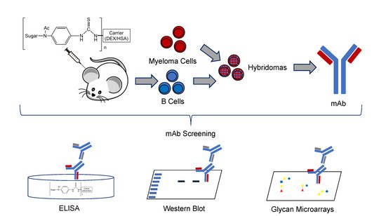
Monoclonal antibodies (mAbs) that recognize glycans are useful tools to assess carbohydrates’ structure and function. We sought to produce IgG mAbs to the human milk oligosaccharide (HMO), lacto-N-fucopentaose III (LNFPIII). LNFPIII contains the Lewisx antigen, which is found on the surface of schistosome parasites. mAbs binding the Lewisx antigen are well-reported in the literature, but mAbs recognizing HMO structures are rare. To generate mAbs, mice were immunized with LNFPIII-DEX (P3DEX) plus CpGs in VacSIM®, a novel vaccine/drug delivery platform. Mice were boosted with LNFPIII-HSA (P3HSA) plus CpGs in Incomplete Freund’s Adjuvant (IFA). Splenocytes from immunized mice were used to generate hybridomas and were screened against LNFPIII conjugates via enzyme-linked immunosorbent assay (ELISA). Three positive hybridomas were expanded, and one hybridoma, producing IgG and IgM antibodies, was cloned via flow cytometry. Clone F1P2H4D8D5 was selected because it produced IgG1 mAbs, but rescreening unexpectedly showed binding to both LNFPIII and lacto-N-neotetraose (LNnT) conjugates. To further assess the specificity of the mAb, we screened it on two glycan microarrays and found no significant binding. This finding suggests that the mAb binds to the acetylphenylenediamine (APD) linker-spacer structure of the conjugate. We present the results herein, suggesting that our new mAb could be a useful probe for conjugates using similar linker spacer structures.

Jessica Ramadhin, Vanessa Silva-Moraes, Thomas Norberg, Donald Harn. Antibodies 20209, 48. https://doi.org/10.3390/antib9030048

Neurochemical and neuroinflammatory perturbations in two Gulf War Illness models: Modulation by the immunotherapeutic LNFPIII

Gulf War Illness (GWI) manifests a multitude of symptoms, including neurological and immunological, and approximately a third of the 1990–1991 Gulf War (GW) veterans suffer from it. This study sought to characterize the acute neurochemical (monoamine) and neuroinflammatory profiles of two established GWI animal models and examine the potential modulatory effects of the novel immunotherapeutic Lacto-N-fucopentaose III (LNFPIII). In Model 1, male C57BL/6 J mice were treated for 10 days with pyridostigmine bromide (PB) and permethrin (PM). In Model 2, a separate cohort of mice were treated for 14 days with PB and N,N-Diethyl-methylbenzamide (DEET), plus corticosterone (CORT) via drinking water on days 8–14 and diisopropylfluorophosphate (DFP) on day 15. LNFPIII was administered concurrently with GWI chemicals treatments. Brain and spleen monoamines and hippocampal inflammatory marker expression were examined by, respectively, HPLC-ECD and qPCR, 6 h post treatment cessation. Serotonergic (5-HT) and dopaminergic (DA) dyshomeostasis caused by GWI chemicals was apparent in multiple brain regions, primarily in the nucleus accumbens (5-HT) and hippocampus (5-HT, DA) for both models. Splenic levels of 5-HT (both models) and norepinephrine (Model 2) were also disrupted by GWI chemicals. LNFPIII treatment prevented many of the GWI chemicals induced monoamine alterations. Hippocampal inflammatory cytokines were increased in both models, but the magnitude and spread of inflammation was greater in Model 2; LNFPIII was anti-inflammatory, more so in the apparently milder Model 1. Overall, in both models, GWI chemicals led to monoamine disbalance and neuroinflammation. LNFPIII co-treatment prevented many of these disruptions in both models, which is indicative of its promise as a potential GWI therapeutic.

J.M. Carpenter, H.E. Gordon, H.D. Ludwig, J.J. Wagner, D.A. Harn, T. Norberg, N.M. Filipov. Neurotoxicology. 2019 Dec 19;77:40-50. doi: 10.1016/j.neuro.2019.12.012