Generic selectors
Exact matches only
Search in title
Search in content
Post Type Selectors

Tag: Chet Joyner

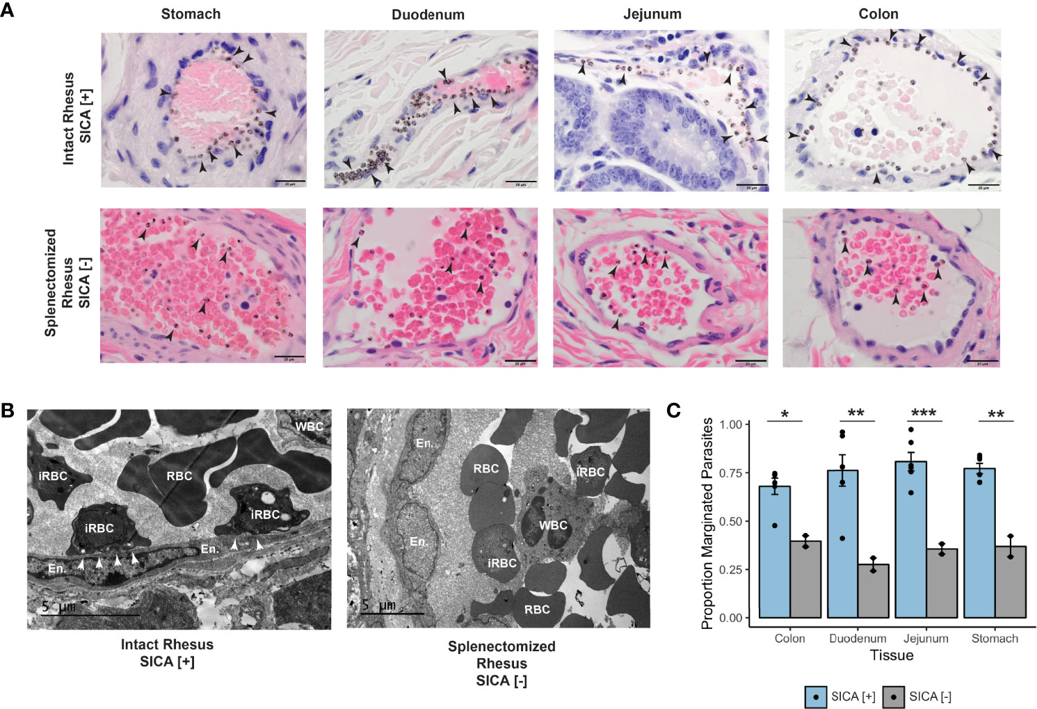
Malaria disrupts the rhesus macaque gut microbiome

Previous studies have suggested that a relationship exists between severity and transmissibility of malaria and variations in the gut microbiome, yet only limited information exists on the temporal dynamics of the gut microbial community during a malarial infection. Here, using a rhesus macaque model of relapsing malaria, we investigate how malaria affects the gut microbiome. In this study, we performed 16S sequencing on DNA isolated from rectal swabs of rhesus macaques over the course of an experimental malarial infection with Plasmodium cynomolgi and analyzed gut bacterial taxa abundance across primary and relapsing infections. We also performed metabolomics on blood plasma from the animals at the same timepoints and investigated changes in metabolic pathways over time. Members of Proteobacteria (family Helicobacteraceae) increased dramatically in relative abundance in the animal’s gut microbiome during peak infection while Firmicutes (family Lactobacillaceae and Ruminococcaceae), Bacteroidetes (family Prevotellaceae) and Spirochaetes amongst others decreased compared to baseline levels. Alpha diversity metrics indicated decreased microbiome diversity at the peak of parasitemia, followed by restoration of diversity post-treatment. Comparison with healthy subjects suggested that the rectal microbiome during acute malaria is enriched with commensal bacteria typically found in the healthy animal’s mucosa. Significant changes in the tryptophan-kynurenine immunomodulatory pathway were detected at peak infection with P. cynomolgi, a finding that has been described previously in the context of P. vivax infections in humans. During relapses, which have been shown to be associated with less inflammation and clinical severity, we observed minimal disruption to the gut microbiome, despite parasites being present. Altogether, these data suggest that the metabolic shift occurring during acute infection is associated with a concomitant shift in the gut microbiome, which is reversed post-treatment.

Danielle N Farinella, Sukhpreet Kaur, ViLinh Tran, Monica Cabrera-Mora, Chester J Joyner, Stacey A Lapp, Suman B Pakala, Mustafa V Nural, Jeremy D DeBarry, Jessica C Kissinger, Dean P Jones, Alberto Moreno, Mary R Galinski, Regina Joice Cordy. Front Cell Infect Microbiol. 2023 Jan 13;12:1058926. doi: 10.3389/fcimb.2022.1058926. eCollection 2022.

MaHPIC malaria systems biology data from Plasmodium cynomolgi sporozoite longitudinal infections in macaques

Plasmodium cynomolgi causes zoonotic malarial infections in Southeast Asia and this parasite species is important as a model for Plasmodium vivax and Plasmodium ovale. Each of these species produces hypnozoites in the liver, which can cause relapsing infections in the blood. Here we present methods and data generated from iterative longitudinal systems biology infection experiments designed and performed by the Malaria Host-Pathogen Interaction Center (MaHPIC) to delve deeper into the biology, pathogenesis, and immune responses of P. cynomolgi in the Macaca mulatta host. Infections were initiated by sporozoite inoculation. Blood and bone marrow samples were collected at defined timepoints for biological and computational experiments and integrative analyses revolving around primary illness, relapse illness, and subsequent disease and immune response patterns. Parasitological, clinical, haematological, immune response, and -omic datasets (transcriptomics, proteomics, metabolomics, and lipidomics) including metadata and computational results have been deposited in public repositories. The scope and depth of these datasets are unprecedented in studies of malaria, and they are projected to be a F.A.I.R., reliable data resource for decades.

Jeremy D DeBarry, Mustafa V Nural, Suman B Pakala, Vishal Nayak, Susanne Warrenfeltz, Jay Humphrey, Stacey A Lapp, Monica Cabrera-Mora, Cristiana F A Brito, Jianlin Jiang, Celia L Saney, Allison Hankus, Hannah M Stealey, Megan B DeBarry, Nicolas Lackman, Noah Legall, Kevin Lee, Yan Tang, Anuj Gupta, Elizabeth D Trippe, Robert R Bridger, Daniel Brent Weatherly, Mariko S Peterson, Xuntian Jiang, ViLinh Tran, Karan Uppal, Luis L Fonseca, Chester J Joyner, Ebru Karpuzoglu, Regina J Cordy, Esmeralda V S Meyer, Lance L Wells, Daniel S Ory, F Eun-Hyung Lee, Rabindra Tirouvanziam, Juan B Gutiérrez 1, Chris Ibegbu, Tracey J Lamb, Jan Pohl, Sarah T Pruett, Dean P Jones, Mark P Styczynski, Eberhard O Voit, Alberto Moreno, Mary R Galinski, Jessica C Kissinger. Sci Data. 2022 Nov 24;9(1):722. doi: 10.1038/s41597-022-01755-y.

Single-cell RNA profiling of Plasmodium vivax-infected hepatocytes reveals parasite- and host- specific transcriptomic signatures and therapeutic targets

The resilience of Plasmodium vivax, the most widely-distributed malaria-causing parasite in humans, is attributed to its ability to produce dormant liver forms known as hypnozoites, which can activate weeks, months, or even years after an initial mosquito bite. The factors underlying hypnozoite formation and activation are poorly understood, as is the parasite’s influence on the host hepatocyte. Here, we shed light on transcriptome-wide signatures of both the parasite and the infected host cell by sequencing over 1,000 P. vivax-infected hepatocytes at single-cell resolution. We distinguish between replicating schizonts and hypnozoites at the transcriptional level, identifying key differences in transcripts encoding for RNA-binding proteins associated with cell fate. In infected hepatocytes, we show that genes associated with energy metabolism and antioxidant stress response are upregulated, and those involved in the host immune response downregulated, suggesting both schizonts and hypnozoites alter the host intracellular environment. The transcriptional markers in schizonts, hypnozoites, and infected hepatocytes revealed here pinpoint potential factors underlying dormancy and can inform therapeutic targets against P. vivax liver-stage infection.

Anthony A Ruberto, Steven P Maher, Amélie Vantaux, Chester J Joyner, Caitlin Bourke, Balu Balan, Aaron Jex, Ivo Mueller, Benoit Witkowski, Dennis E Kyle. Front Cell Infect Microbiol. 2022 Aug 25;12:986314. doi: 10.3389/fcimb.2022.986314. eCollection 2022.

Plasmodium knowlesi Cytoadhesion Involves SICA Variant Proteins

Plasmodium knowlesi poses a health threat throughout Southeast Asian communities and currently causes most cases of malaria in Malaysia. This zoonotic parasite species has been studied in Macaca mulatta (rhesus monkeys) as a model for severe malarial infections, chronicity, and antigenic variation. The phenomenon of Plasmodium antigenic variation was first recognized during rhesus monkey infections. Plasmodium-encoded variant proteins were first discovered in this species and found to be expressed at the surface of infected erythrocytes, and then named the Schizont-Infected Cell Agglutination (SICA) antigens. SICA expression was shown to be spleen dependent, as SICA expression is lost after P. knowlesi is passaged in splenectomized rhesus. Here we present data from longitudinal P. knowlesi infections in rhesus with the most comprehensive analysis to date of clinical parameters and infected red blood cell sequestration in the vasculature of tissues from 22 organs. Based on the histopathological analysis of 22 tissue types from 11 rhesus monkeys, we show a comparative distribution of parasitized erythrocytes and the degree of margination of the infected erythrocytes with the endothelium. Interestingly, there was a significantly higher burden of parasites in the gastrointestinal tissues, and extensive margination of the parasites along the endothelium, which may help explain gastrointestinal symptoms frequently reported by patients with P. knowlesi malarial infections. Moreover, this margination was not observed in splenectomized rhesus that were infected with parasites not expressing the SICA proteins. This work provides data that directly supports the view that a subpopulation of P. knowlesi parasites cytoadheres and sequesters, likely via SICA variant antigens acting as ligands. This process is akin to the cytoadhesive function of the related variant antigen proteins, namely Erythrocyte Membrane Protein-1, expressed by Plasmodium falciparum.

Mariko S Peterson, Chester J Joyner, Stacey A Lapp, Jessica A Brady, Jennifer S Wood, Monica Cabrera-Mora, Celia L Saney, Luis L Fonseca, Wayne T Cheng, Jianlin Jiang, Stephanie R Soderberg, Mustafa V Nural, Allison Hankus, Deepa Machiah, Ebru Karpuzoglu, Jeremy D DeBarry, Rabindra Tirouvanziam, Jessica C Kissinger, Alberto Moreno, Sanjeev Gumber, Eberhard O Voit, Juan B Gutierrez, Regina Joice Cordy, Mary R Galinski. Front Cell Infect Microbiol. 2022 Jun 23;12:888496. doi: 10.3389/fcimb.2022.888496. eCollection 2022.

Clinical recovery of Macaca fascicularis infected with Plasmodium knowlesi

Background: Kra monkeys (Macaca fascicularis), a natural host of Plasmodium knowlesi, control parasitaemia caused by this parasite species and escape death without treatment. Knowledge of the disease progression and resilience in kra monkeys will aid the effective use of this species to study mechanisms of resilience to malaria. This longitudinal study aimed to define clinical, physiological and pathological changes in kra monkeys infected with P. knowlesi, which could explain their resilient phenotype.

Methods: Kra monkeys (n = 15, male, young adults) were infected intravenously with cryopreserved P. knowlesi sporozoites and the resulting parasitaemias were monitored daily. Complete blood counts, reticulocyte counts, blood chemistry and physiological telemetry data (n = 7) were acquired as described prior to infection to establish baseline values and then daily after inoculation for up to 50 days. Bone marrow aspirates, plasma samples, and 22 tissue samples were collected at specific time points to evaluate longitudinal clinical, physiological and pathological effects of P. knowlesi infections during acute and chronic infections.

Results: As expected, the kra monkeys controlled acute infections and remained with low-level, persistent parasitaemias without anti-malarial intervention. Unexpectedly, early in the infection, fevers developed, which ultimately returned to baseline, as well as mild to moderate thrombocytopenia, and moderate to severe anaemia. Mathematical modelling and the reticulocyte production index indicated that the anaemia was largely due to the removal of uninfected erythrocytes and not impaired production of erythrocytes. Mild tissue damage was observed, and tissue parasite load was associated with tissue damage even though parasite accumulation in the tissues was generally low.

Conclusions: Kra monkeys experimentally infected with P. knowlesi sporozoites presented with multiple clinical signs of malaria that varied in severity among individuals. Overall, the animals shared common mechanisms of resilience characterized by controlling parasitaemia 3-5 days after patency, and controlling fever, coupled with physiological and bone marrow responses to compensate for anaemia. Together, these responses likely minimized tissue damage while supporting the establishment of chronic infections, which may be important for transmission in natural endemic settings. These results provide new foundational insights into malaria pathogenesis and resilience in kra monkeys, which may improve understanding of human infections.

Mariko S Peterson, Chester J Joyner, Jessica A Brady, Jennifer S Wood, Monica Cabrera-Mora, Celia L Saney, Luis L Fonseca, Wayne T Cheng, Jianlin Jiang, Stacey A Lapp, Stephanie R Soderberg, Mustafa V Nural, Jay C Humphrey, Allison Hankus, Deepa Machiah, Ebru Karpuzoglu, Jeremy D DeBarry, MaHPIC-Consortium; Rabindra Tirouvanziam, Jessica C Kissinger, Alberto Moreno, Sanjeev Gumber, Eberhard O Voit, Juan B Gutiérrez, Regina Joice Cordy, Mary R Galinski. Clinical recovery of Macaca fascicularis infected with Plasmodium knowlesiMalar J 20, 486 (2021). https://doi.org/10.1186/s12936-021-03925-6

Generation of human long-lived plasma cells by developmentally regulated epigenetic imprinting

Antibody secreting cells (ASCs) circulate after vaccination and infection and migrate to the BM where a subset known as long-lived plasma cells (LLPCs) persists and secrete antibodies for a lifetime. The mechanisms by which circulating ASCs become LLPCs are not well elucidated. Here, we show that human blood ASCs have distinct morphology, transcriptomes, and epigenetics compared with BM LLPCs. Compared with blood ASCs, BM LLPCs have decreased nucleus/cytoplasm ratio but increased endoplasmic reticulum and numbers of mitochondria. LLPCs up-regulate pro-survival genes MCL1BCL2, and BCL-XL while simultaneously down-regulating pro-apoptotic genes HRK1CASP3, and CASP8 Consistent with reduced gene expression, the pro-apoptotic gene loci are less accessible in LLPCs. Of the pro-survival genes, only BCL2 is concordant in gene up-regulation and loci accessibility. Using a novel in vitro human BM mimetic, we show that blood ASCs undergo similar morphological and molecular changes that resemble ex vivo BM LLPCs. Overall, our study demonstrates that early-minted blood ASCs in the BM microniche must undergo morphological, transcriptional, and epigenetic changes to mature into apoptotic-resistant LLPCs.

Chester J Joyner, Ariel M Ley, Doan C Nguyen, Mohammad Ali, Alessia Corrado, Christopher Tipton, Christopher D Scharer, Tian Mi, Matthew C Woodruff, Jennifer Hom, Jeremy M Boss, Meixue Duan, Greg Gibson, Danielle Roberts, Joel Andrews, Sagar Lonial, Inaki Sanz, F Eun-Hyung Lee. Life Sci Alliance. 2021 Dec 24;5(3):e202101285. doi: 10.26508/lsa.202101285.

A Phenotypic Screen for the Liver Stages of Plasmodium vivax

Control of malaria caused by Plasmodium vivax can be improved by the discovery and development of novel drugs against the parasite’s liver stage, which includes relapse-causing hypnozoites. Several recent reports describe breakthroughs in the culture of the P. vivax liver stage in 384-well microtiter plates, with the goal of enabling a hypnozoite-focused drug screen. Herein we describe assay details, protocol developments, and different assay formats to interrogate the chemical sensitivity of the P. vivax liver stage in one such medium-throughput platform. The general assay protocol includes seeding of primary human hepatocytes which are infected with P. vivax sporozoites generated from the feeding of Anopheles dirus mosquitoes on patient isolate bloodmeals. This protocol is unique in that, after source drug plates are supplied, all culture-work steps have been optimized to preclude the need for automated liquid handling, thereby allowing the assay to be performed within resource-limited laboratories in malaria-endemic countries. Throughput is enhanced as complex culture methods, such as extracellular matrix overlays, multiple cell types in co-culture, or hepatic spheroids, are excluded as the workflow consists entirely of routine culture methods for adherent cells. Furthermore, installation of a high-content imager at the study site enables assay data to be read and transmitted with minimal logistical delays. Herein we detail distinct assay improvements which increase data quality, provide a means to limit the confounding effect of hepatic metabolism on assay data, and detect activity of compounds with a slow-clearance phenotype.

Steven P. Maher, Amélie Vantaux, Caitlin A. Cooper, Nathan M. Chasen, Wayne T. Cheng, Chester J. Joyner, Roman Manetsch, Benoît Witkowski, Dennis Kyle. 2021. Bio-Protocol. 11(23): DOI: 10.21769/BioProtoc.4253

UGA researchers developing new models for malaria drug development and testing

Chet Joyner and Dennis Kyle
CTEGD member Chet Joyner and CTEGD director Dennis Kyle receive a grant from the Bill & Melinda Gates Foundation for malaria drug development and testing

Two UGA researchers are working to make it easier to develop effective treatments for malaria, a disease that sickens millions worldwide and kills hundreds of thousands each year.

In tropical climates around the globe, malaria poses a grave risk to already vulnerable populations. In 2019, the World Health Organization estimated that there were 229 million clinical cases of malaria worldwide and 409,000 deaths, usually in children below the age of five.

Currently, developing and testing drugs for malaria requires scientists to work in areas where the disease is prevalent or to work with expensive, hard-to-source equipment.  Chester Joyner, an Assistant Professor in the Center for Vaccines and Immunology, and Dennis Kyle, Professor of Infectious Diseases and Cellular Biology, are working to reduce those barriers to malaria drug testing and development.

Joyner and Kyle aim to establish systems that rely on equipment most researchers can obtain: a petri dish. If successful, Joyner says this new culture system will reduce costs and be distributed more easily to advance drug and vaccine research. The University of Georgia College of Veterinary Medicine received a grant for malaria drug development and testing from the Bill & Melinda Gates Foundation.

Worldwide, there are many malaria-causing parasites that result in varying degrees of illness. Joyner and Kyle’s research focuses on defeating one of the most challenging: Plasmodium vivax. Unlike many other malaria parasites, P. vivax can lie dormant in the livers of its hosts—allowing the infected to travel abroad completely unaware that they’re carrying a potentially deadly passenger.

“Most infections with P. vivax are not due to new infections,” says Joyner. “These infections come from this parasite activating and potentially causing disease and sustaining transmission.”

Malaria disproportionately affects the poorest communities in the world, creating a cycle of disease and poverty that current treatments have improved but been unable to stop. However, treating the dormant forms of P. vivax has been particularly challenging because they can cause more harm than good in at-risk populations like pregnant women and people with certain blood conditions.

“We want researchers to have access to technologies to study P. vivax and develop new approaches to control and eliminate this parasite,” Joyner explains.


This article first appeared at https://give.uga.edu/uga-researchers-developing-new-models-for-malaria-drug-development-and-testing/

Acute Plasmodium Infection Promotes Interferon-Gamma-Dependent Resistance to Ebola Virus Infection

During the 2013-2016 Ebola virus (EBOV) epidemic, a significant number of patients admitted to Ebola treatment units were co-infected with Plasmodium falciparum, a predominant agent of malaria. However, there is no consensus on how malaria impacts EBOV infection. The effect of acute Plasmodium infection on EBOV challenge was investigated using mouse-adapted EBOV and a biosafety level 2 (BSL-2) model virus. We demonstrate that acute Plasmodium infection protects from lethal viral challenge, dependent upon interferon gamma (IFN-γ) elicited as a result of parasite infection. Plasmodium-infected mice lacking the IFN-γ receptor are not protected. Ex vivo incubation of naive human or mouse macrophages with sera from acutely parasitemic rodents or macaques programs a proinflammatory phenotype dependent on IFN-γ and renders cells resistant to EBOV infection. We conclude that acute Plasmodium infection can safeguard against EBOV by the production of protective IFN-γ. These findings have implications for anti-malaria therapies administered during episodic EBOV outbreaks in Africa.

Kai J Rogers, Olena Shtanko, Rahul Vijay, Laura N Mallinger, Chester J Joyner, Mary R Galinski, Noah S Butler, Wendy Maury. Cell Rep. 2020 Mar 24;30(12):4041-4051.e4. doi: 10.1016/j.celrep.2020.02.104.