Generic selectors
Exact matches only
Search in title
Search in content
Post Type Selectors

Tag: calcium

Regulation of Calcium entry by cyclic GMP signaling in Toxoplasma gondii
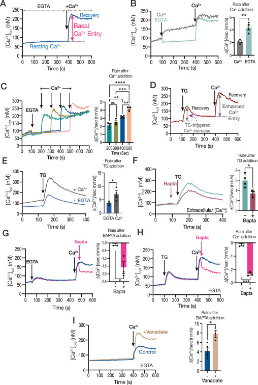
Figure 1. Calcium entry through the plasma membrane of extracellular T. gondii tachyzoites.


Ca2+ signaling impacts almost every aspect of cellular life. Ca2+ signals are generated through the opening of ion channels that permit the flow of Ca2+ down an electrochemical gradient. Cytosolic Ca2+ fluctuations can be generated through Ca2+ entry from the extracellular milieu or release from intracellular stores. In Toxoplasma gondii, Ca2+ ions play critical roles in several essential functions for the parasite like invasion of host cells, motility and egress. Plasma membrane Ca2+ entry in T. gondii was previously shown to be activated by cytosolic calcium and inhibited by the voltage-operated Ca2+ channel blocker nifedipine. However, Ca2+ entry in T. gondii did not show the classical characteristics of store regulation. In this work, we characterized the mechanism by which cytosolic Ca2+ regulates plasma membrane Ca2+ entry in extracellular T. gondii tachyzoites loaded with the Ca2+ indicator Fura 2. We compared the inhibition by nifedipine with the effect of the broad spectrum TRP channel inhibitor, anthranilic acid or ACA and we find that both inhibitors act on different Ca2+ entry activities. We demonstrate, using pharmacological and genetic tools, that an intracellular signaling pathway engaging cyclicGMP (cGMP), protein kinase G (PKG), Ca2+ and the phosphatidyl inositol phospholipase C (PI-PLC) affects Ca2+ entry and we present a model for crosstalk between cGMP and cytosolic Ca2+ for the activation of T. gondii‘s lytic cycle traits.

Miryam A Hortua Triana, Karla M Márquez-Nogueras, Mojtaba Sedigh Fazli, Shannon Quinn, Silvia N J Moreno. J Biol Chem. 2024 Feb 19:105771. doi: 10.1016/j.jbc.2024.105771

Advances in the cellular biology, biochemistry, and molecular biology of acidocalcisomes

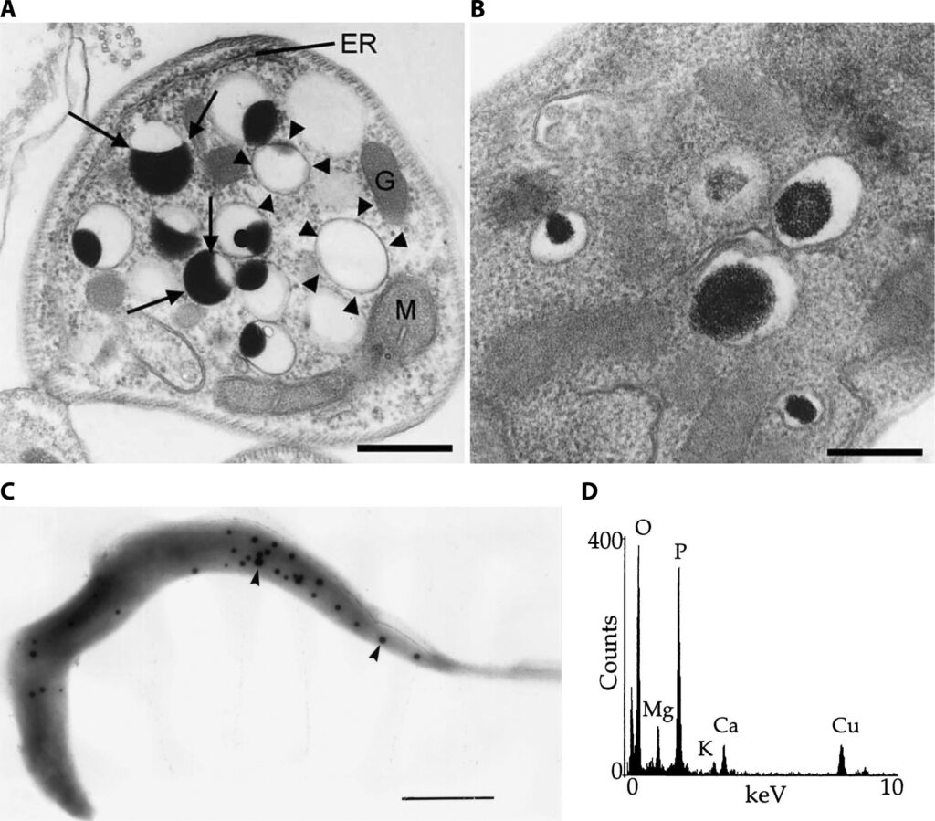
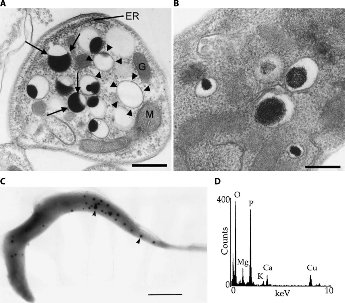
Fig 1 Ultrastructure of acidocalcisomes
Fig 1 Ultrastructure of acidocalcisomes.

Acidocalcisomes are organelles conserved during evolution and closely related to the so-called volutin granules of bacteria and archaea, to the acidocalcisome-like vacuoles of yeasts, and to the lysosome-related organelles of animal species. All these organelles have in common their acidity and high content of polyphosphate and calcium. They are characterized by a variety of functions from storage of phosphorus and calcium to roles in Ca2+ signaling, osmoregulation, blood coagulation, and inflammation. They interact with other organelles through membrane contact sites or by fusion, and have several enzymes, pumps, transporters, and channels.

Roberto Docampo. Microbiol Mol Biol Rev. 2023 Dec 15:e0004223. doi: 10.1128/mmbr.00042-23.

Praziquantel target validation of a Ca2+ permeable channel in schistosomes

Highlights

  • Schistosomiasis is a devastating neglected helminthic disease.
  • Praziquantel (PZQ) is the most important drug against schistosomiasis.
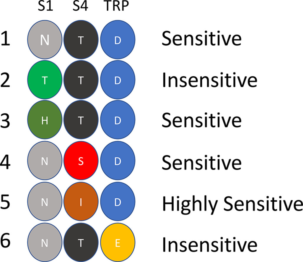
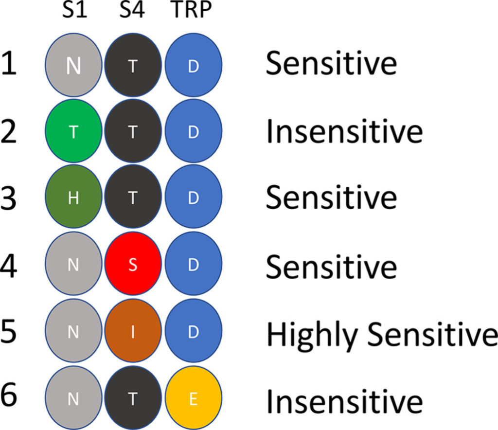
  • Previous work identified a TRP channel of the melastatin type as a PZQ target.
  • Molecular studies now reveal the basis for varied PZQ sensitivity of different helminths.

Roberto Docampo. Cell Calcium. 2023 Jan 18;110:102698. doi: 10.1016/j.ceca.2023.102698

Calcium signaling in intracellular protist parasites

Calcium ion (Ca2+) signaling is one of the most frequently employed mechanisms of signal transduction by eukaryotic cells, and starts with either Ca2+ release from intracellular stores or Ca2+ entry through the plasma membrane. In intracellular protist parasites Ca2+ signaling initiates a sequence of events that may facilitate their invasion of host cells, respond to environmental changes within the host, or regulate the function of their intracellular organelles. In this review we examine recent findings in Ca2+ signaling in two groups of intracellular protist parasites that have been studied in more detail, the apicomplexan and the trypanosomatid parasites.

Roberto Docampo, Silvia Nj Moreno. Current Opinion in Microbiology 2021, 64:33–40. https://doi.org/10.1016/j.mib.2021.09.002

Ca2+ entry at the plasma membrane and uptake by acidic stores is regulated by the activity of the V‐H+‐ATPase in Toxoplasma gondii

Ca2+ is a universal intracellular signal that regulates many cellular functions. In Toxoplasma gondii, the controlled influx of extracellular and intracellular Ca2+ into the cytosol initiates a signaling cascade that promotes pathogenic processes like tissue destruction and dissemination. In this work we studied the role of proton transport in cytosolic Ca2+ homeostasis and the initiation of Ca2+ signaling. We used a T. gondii mutant of the V-ATPase, a pump previously shown to transport protons to the extracellular medium, control intracellular pH and membrane potential and we show that proton gradients are important for maintaining resting cytosolic Ca2+ at physiological levels and for Ca2+ influx. Proton transport was also important for Ca2+ storage by acidic stores and, unexpectedly, the endoplasmic reticulum. Proton transport impacted the amount of polyphosphate (polyP), a phosphate polymer that binds Ca2+ and concentrate in acidocalcisomes. This was supported by the co-localization of the vacuolar transporter chaperone 4 (VTC4), the catalytic subunit of the VTC complex that synthesizes polyP, with the V-ATPase in acidocalcisomes. Our work show that proton transport regulate plasma membrane Ca2+ transport and control acidocalcisome polyP and Ca2+ content impacting Ca2+ signaling and downstream stimulation of motility and egress in T. gondii.

Andrew J Stasic, Eric J Dykes, Ciro D Cordeiro, Stephen A Vella, Mojtaba S Fazli, Shannon Quinn, Roberto Docampo, Silvia N J Moreno. Mol Microbiol. 2021 Apr 1. doi: 10.1111/mmi.14722

RGS10 physically and functionally interacts with STIM2 and requires store-operated calcium entry to regulate proinflammatory gene expression in microglia

Chronic activation of microglia is a driving factor in the progression of neuroinflammatory diseases, and mechanisms that regulate microglial inflammatory signaling are potential targets for novel therapeutics. Regulator of G protein Signaling 10 is the most abundant RGS protein in microglia, where it suppresses inflammatory gene expression and reduces microglia-mediated neurotoxicity. In particular, microglial RGS10 downregulates the expression of pro-inflammatory mediators including cyclooxygenase 2 (COX-2) following stimulation with lipopolysaccharide (LPS). However, the mechanism by which RGS10 affects inflammatory signaling is unknown and is independent of its canonical G protein targeted mechanism. Here, we sought to identify non-canonical RGS10 interacting partners that mediate its anti-inflammatory mechanism. Through RGS10 co-immunoprecipitation coupled with mass spectrometry, we identified STIM2, an endoplasmic reticulum (ER) localized calcium sensor and a component of the store-operated calcium entry (SOCE) machinery, as a novel RGS10 interacting protein in microglia. Direct immunoprecipitation experiments confirmed RGS10-STIM2 interaction in multiple microglia and macrophage cell lines, as well as in primary cells, with no interaction observed with the homologue STIM1. We further determined that STIM2, Orai channels, and the Ca2+--dependent phosphatase calcineurin are essential for LPS-induced COX-2 production in microglia, and this pathway is required for the inhibitory effect of RGS10 on COX-2. Additionally, our data demonstrated that RGS10 suppresses SOCE triggered by ER calcium depletion and that ER calcium depletion, which induces SOCE, amplifies proinflammatory genes. In addition to COX-2, we also show that RGS10 suppresses the expression of proinflammatory cytokines in microglia in response to thrombin and LPS stimulation, and all of these effects require SOCE. Collectively, the physical and functional links between RGS10 and STIM2 suggest a complex regulatory network connecting RGS10, SOCE, and pro-inflammatory gene expression in microglia, with broad implications in the pathogenesis and treatment of chronic neuroinflammation.

Menbere Wendimu, Mohammed Alqinyah, Stephen Vella, Phillip Dean, Faris Almutairi, Roseanne Davila Rivera, Shima Rayatpisheh, James Wohlschlegel, Silvia Moreno, Shelley B Hooks. Cell Signal. 2021 Mar 8;109974. doi: 10.1016/j.cellsig.2021.109974

IP3 receptor-mediated Ca2+ release from acidocalcisomes regulates mitochondrial bioenergetics and prevents autophagy in Trypanosoma cruzi

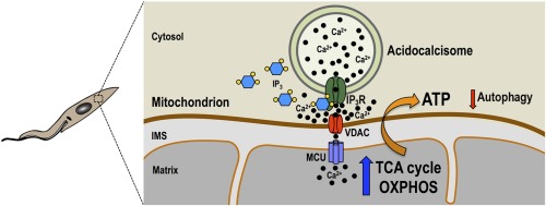
In contrast to animal cells, the inositol 1,4,5-trisphosphate receptor of Trypanosoma cruzi (TcIP3R) localizes to acidocalcisomes instead of the endoplasmic reticulum. Here, we present evidence that TcIP3R is a Ca2+ release channel gated by IP3 when expressed in DT40 cells knockout for all vertebrate IP3 receptors, and is required for Ca2+ uptake by T. cruzi mitochondria, regulating pyruvate dehydrogenase dephosphorylation and mitochondrial O2 consumption, and preventing autophagy. Localization studies revealed its co-localization with an acidocalcisome marker in all life cycle stages of the parasite. Ablation of TcIP3R by CRISPR/Cas9 genome editing caused: a) a reduction in O2 consumption rate and citrate synthase activity; b) decreased mitochondrial Ca2+ transport without affecting the membrane potential; c) increased ammonia production and AMP/ATP ratio; d) stimulation of autophagosome formation, and e) marked defects in growth of culture forms (epimastigotes) and invasion of host cells by infective stages (trypomastigotes). Moreover, TcIP3R overexpressing parasites showed decreased metacyclogenesis, trypomastigote host cell invasion and intracellular amastigote replication. In conclusion, the results suggest a modulatory activity of TcIP3R-mediated acidocalcisome Ca2+ release on cell bioenergetics in T. cruzi.

Miguel A. Chiurillo, Noelia Lander, Anibal E. Vercesi, Roberto Docampo, 2020. Cell Calcium; 92:102284, https://doi.org/10.1016/j.ceca.2020.102284.

Genetic Indicators for Calcium Signaling Studies in Toxoplasma gondii

Fluctuations of the cytosolic calcium ion (Ca2+) concentration regulate a variety of cellular functions in all eukaryotes. Cells express a sophisticated set of mechanisms to balance the cytosolic Ca2+ levels and the signals that elevate Ca2+ in the cytosol are compensated by mechanisms that reduce it. Alterations in Ca2+-dependent homeostatic mechanisms are the cause of many prominent diseases in humans, such as heart failure or neuronal death.

The genetic tractability of Toxoplasma gondii and the availability of genetic tools enabled the use of Genetically Encoded Calcium Indicators (GECIs) expressed in the cytoplasm, which started a new era in the studies of Toxoplasma calcium signaling. It was finally possible to see Ca2+ oscillations prior to exit of the parasite from host cells. Years after Endo et al showed that ionophores triggered egress, the assumption that oscillations occur prior to egress from host cells has been validated by experiments using GECIs. GECIs allowed the visualization of specific Ca2+ signals in live intracellular parasites and to distinguish these signals from host cell calcium fluctuations. In this chapter we present an overview describing “tried and true” methods of our lab who pioneered the first use of GECI’s in Toxoplasma, including GECI choice, methodology for transfection and selection of ideal clones, their characterization, and the use of GECI-expressing parasites for fluorometric and microscopic analysis.

Stephen A. Vella, Abigail Calixto, Beejan Asady, Zhu-Hong Li, Silvia N. J. Moreno. Methods Mol Biol. 2020;2071:187-207. doi: 10.1007/978-1-4939-9857-9_11.

Trainee Spotlight: Stephen Vella

Stephen Vella is a Ph.D. trainee in Silvia Moreno’s laboratory. He is originally from Indiana where he received his B.S. in microbiology at Indiana University. In his first year at UGA, he was awarded an Excellence in Graduate Recruitment Award and a Provost’s Scholars of Excellence Award Fellowship. He has also been awarded an Outstanding Poster Presentation at the Molecular Parasitology Meeting in 2016. And in 2017, he was awarded a T32 fellowship from CTEGD.