Generic selectors
Exact matches only
Search in title
Search in content
Post Type Selectors

Tag: Brugia malayi

An Overview of Management Considerations for Mongolian Gerbils (Meriones unguiculatus), Cats (Felis catus), and Dogs (Canis familiaris) as Hosts for Brugia Infection

Lymphatic filariasis is a mosquito-borne parasitic infection affecting an estimated 51.4 million people. Brugia malayi and Brugia pahangi are used in research because common nonprimate research species such as Mongolian gerbils (Meriones unguiculatus), cats (Felis catus), and dogs (Canis familiaris) can maintain the life cycle of these species of filarial nematodes. Although overall care and management of animals infected with Brugia spp. is relatively straightforward, there are some unique challenges and special considerations that must be addressed when managing a research colony infected with these parasites. In this review, we discuss our experience, share insight into biosafety and clinical management, and describe the expected clinical signs associated with Brugia infection in gerbils, cats, and dogs.

Catherine A Chambers, Christopher C Evans, Gianni A Campellone, Mary A McCrackin, Andrew R Moorhead, Leanne C Alworth. Comp Med. 2024 Jun 26. doi: 10.30802/AALAS-CM-24-034.

The Domestic Dog as a Laboratory Host for Brugia malayi

Of the three nematodes responsible for lymphatic filariasis in humans, only Brugia malayi is actively maintained in research settings owing to its viability in small animal hosts, principal among which is the domestic cat. While the microfilaremic feline host is necessary for propagation of parasites on any significant scale, this system is plagued by a number of challenges not as pronounced in canine filarial models. For this reason, we investigated the capacity in which dogs may serve as competent laboratory hosts for B. malayi. We infected a total of 20 dogs by subcutaneous injection of 500 B. malayi third-stage larvae (L3) in either a single (n = 10) or repeated infection events (125 L3 per week for four weeks; n = 10). Within each group, half of the individuals were injected in the inguinal region and half in the dorsum of the hind paw. To track the course of microfilaremia in this host, blood samples were examined by microscopy biweekly for two years following infection. Additionally, to identify cellular responses with potential value as predictors of patency, we measured peripheral blood leukocyte counts for the first year of infection. A total of 10 of 20 dogs developed detectable microfilaremia. Peak microfilaria density varied but attained levels useful for parasite propagation (median = 1933 mL-1; range: 33-9950 mL-1). Nine of these dogs remained patent at 104 weeks. A two-way ANOVA revealed no significant differences between infection groups in lifetime microfilaria production (p = 0.42), nor did regression analysis reveal any likely predictive relationships to leukocyte values. The results of this study demonstrate the competence of the dog as a host for B. malayi and its potential to serve in the laboratory role currently provided by the cat, while also clarifying the potential for zoonosis in filariasis-endemic regions.

Christopher C Evans, Katelin E Greenway, Elyssa J Campbell, Michael T Dzimianski, Abdelmoneim Mansour, John W McCall, Andrew R Moorhead. Pathogens. 2022 Sep 21;11(10):1073. doi: 10.3390/pathogens11101073.

Recognition and killing of Brugia malayi microfilariae by human immune cells is dependent on the parasite sample and is not altered by ivermectin treatment

graphical abstract

Abstract

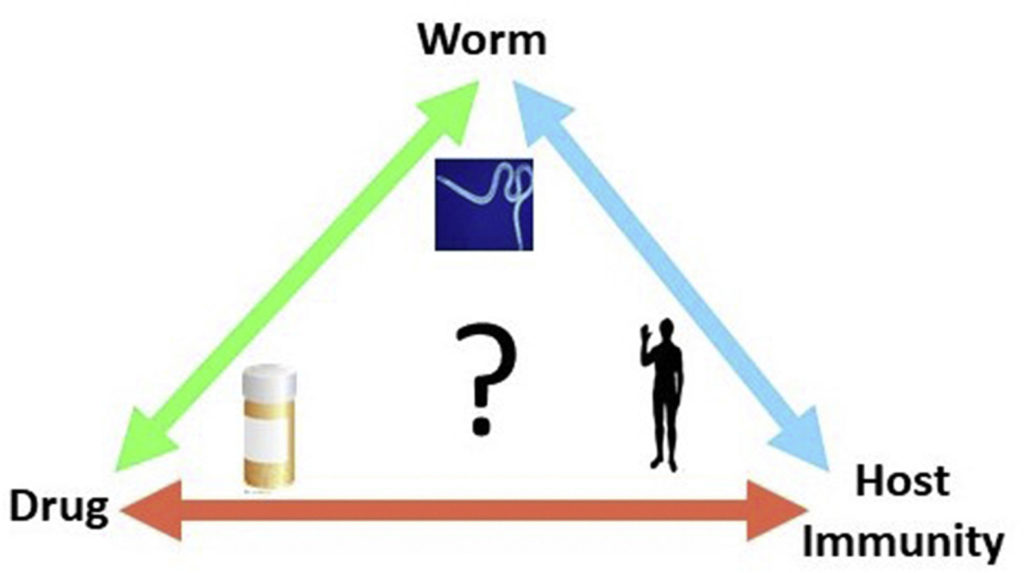
Mass administration of macrocyclic lactones targets the transmission of the causative agents of lymphatic filariasis to their insect vectors by rapidly clearing microfilariae (Mf) from the circulation. It has been proposed that the anti-filarial action of these drugs may be mediated through the host immune system. We recently developed an in vitro assay for monitoring the attachment to and killing of B. malayi Mf by human neutrophils (PMNs) and monocytes (PBMCs), however, the levels of both cell to worm attachment and leukocyte mediated Mf killing varied greatly between individual experiments. To determine whether differences in an individual’s immune cells or the Mf themselves might account for the variability in survival, PMNs and PBMCs were isolated from 12 donors every week for 4 weeks and the cells used for survival assays with a different batch of Mf, thereby keeping donors constant but varying the Mf sample. Results from these experiments indicate that, overall, killing is Mf-rather than donor-dependent. To assess whether ivermectin (IVM) or diethylcarbamazine (DEC) increase killing, Mf were incubated either alone or with immune cells in the presence of IVM or DEC. Neither drug induced a significant difference in the survival of Mf whether cultured with or without cells, with the exception of DEC at 2 h post incubation. In addition, human PBMCs and PMNs were incubated with IVM or DEC for 1 h or 16 h prior to RNA extraction and Illumina sequencing. Although donor-to-donor variation may mask subtle differences in gene expression, principle component analysis of the RNASeq data indicates that there is no significant change in the expression of any genes from the treated cells versus controls. Together these data suggest that IVM and DEC have little direct effect on immune cells involved in the rapid clearance of Mf from the circulation.

Barbara J. Reaves, Connor Wallis, Ciaran J.McCoy, W. Walter Lorenz, Balazs Rada, Adrian J.Wolstenholme. 2018. International Journal for Parasitology: Drugs and Drug Resistance; 6(3): 587-595.