Generic selectors
Exact matches only
Search in title
Search in content
Post Type Selectors

Tag: African Trypansomiasis

Pseudokinase NRP1 facilitates endocytosis of transferrin in the African trypanosome

Trypanosoma brucei causes human African trypanosomiasis (HAT) and nagana in cattle. During infection of a vertebrate, endocytosis of host transferrin (Tf) is important for viability of the parasite. The majority of proteins involved in trypanosome endocytosis of Tf are unknown. Here we identify pseudokinase NRP1 (Tb427tmp.160.4770) as a regulator of Tf endocytosis. Genetic knockdown of NRP1 inhibited endocytosis of Tf without blocking uptake of bovine serum albumin. Binding of Tf to the flagellar pocket was not affected by knockdown of NRP1. However the quantity of Tf per endosome dropped significantly, consistent with NRP1 promoting robust capture and/or retention of Tf in vesicles. NRP1 is involved in motility of Tf-laden vesicles since distances between endosomes and the kinetoplast were reduced after knockdown of the gene. In search of possible mediators of NRP1 modulation of Tf endocytosis, the gene was knocked down and the phosphoproteome analyzed. Phosphorylation of protein kinases forkhead, NEK6, and MAPK10 was altered, in addition to EpsinR, synaptobrevin and other vesicle-associated proteins predicted to be involved in endocytosis. These candidate proteins may link NRP1 functionally either to protein kinases or to vesicle-associated proteins.

Gaurav Kumar, Bryanna Thomas, Kojo Mensa-Wilmot. Sci Rep. 2022 Nov 3;12(1):18572. doi: 10.1038/s41598-022-22054-x.

Trainee Adds New Tool to the Trypanosome Toolbox

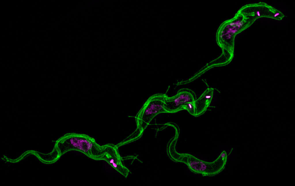
Trypanosoma brucei stained with mCLING
mCLING-staining of membrane (green) and DAPI staining of DNA (magenta) in trypanosomes in various life cycle stages. Flagella appear as tube-like structures along the length of the cell body. Nanotubes can be seen projecting outward from the periphery of the cells.

When Ph.D. trainee Justin Wiedeman started investigating the role of protein kinase TbCK1.2, an enzyme found near the flagellum of Trypanosoma brucei, he quickly ran into a problem common to parasitologists. He needed a better tool for visualizing the membranes of this parasite. Since none of the membrane probes on the market quite did the job, he looked at how he could modify one for his purpose. He found a successful candidate in Synaptic Systems’ mCLING.

What is Trypanosoma brucei?

Trypanosoma brucei is a single cell parasite that causes Human African Trypanosomiasis (HAT), which is also known as African sleeping sickness. HAT occurs in 36 sub-Saharan countries where tsetse flies transmit the parasite to people and livestock. In cattle, the disease is known as nagana. Tsetse fly control efforts have drastically reduced the number of cases. According to the World Health Organization, in 2015, there were around 2,800 cases. However, a person can be infected for months or even years without symptoms. By the time symptoms become evident, the person is in the advanced stages of the disease and their central nervous system is impaired.

New tools are needed to study trypanosomes

There is still much to be learned about the parasite that could lead to better detection and more effective treatment. A major obstacle to the study of this tiny organism is the lack of tools and technology. Kojo Mensa-Wilmot’s research group in the Center for Tropical and Emerging Global Diseases at The University of Georgia has been instrumental in developing techniques and tools to increase the research community’s understanding of T. brucei. Now, Wiedeman has added a new tool to the trypanosome biology toolbox – a general method of outlining trypanosomes in fluorescence microscopy experiments.

“We are the first group to solve this general problem in super-resolution microscopy of T. brucei,” said Wiedeman. “mCLING is a highly versatile tool for studying trypanosome biology – it can be used with live or fixed trypanosomes.”

Fluorescent microscopy has been a leading method of studying T. brucei; however, there are limitations to this technology. Super-resolution microscopy offers great advantages over standard fluorescence microscopy. By employing several techniques to increase resolution, it allows for the observation of objects smaller than what can be seen with visible light.  Yet, it is not without its own limitations, most notably the inability to determine the periphery of cells. Without knowing the outer edges of the parasite, orientation of organelles and other structures within the cell is difficult.

“For Trypanosoma brucei, most of the membrane probes available do not work well in fixed trypanosomes,” said Wiedeman. “Researchers have been forced to use crude methods to outline trypanosomes in fluorescence microscopy.”

These “crude methods” include superimposing a transmitted light image or hand-drawing the outline. However, this workaround only allows for a two-dimensional study of the cell. Therefore, Wiedeman turned to a dye called mCLING that has been developed to track the membranes of neurons using super-resolution microscopy to see if he could adapt the technology to T. brucei membranes.

mCLING allows for the visualization of T. brucei membranes

“mCLING labels the flagellum and plasma membrane vividly, sometimes providing details of cell structure that rivals images obtained with scanning electron microscopy,” said Wiedeman.

Using a combination of standard-resolution and super-resolution fluorescence microscopy, he was able to confirm mCLING labels the plasma and flagellar membranes of T. brucei. Furthermore, using the Zeiss ELYRA S1 super-resolution microscopy in the Biomedical Microscopy Core, mCLING allowed for a 3D reconstruction of the parasite. This is the first time such an image has been reported. Finally, using the new ImageStream X Mark II in the CTEGD Cytometry Shared Resource Laboratory, he discovered mCLING could be used to track endocytosis (the process of importing molecules into the cell) in real time.

Recognizing mCLING’s potential to inform other studies of trypanosome biology, Weideman optimized protocols for using it with immunofluorescence assays and thus making possible what had been impossible with the overlay technique – visualizing the location of organelles in the vertical dimension relative to the cell body.

“It is especially well-suited for studying flagellar membrane biogenesis as well as kinetically tracking uptake of the plasma membrane into vesicles inside trypanosomes,” said Wiedeman. Other laboratories have already implemented these protocols in their own research. Steve Hajduk’s group, also in the Center for Tropical and Emerging Global Diseases, is using mCLING to study nanotubes in T. brucei.

This tool will allow for the study of trypanosomes in finer detail than ever before and the Mensa-Wilmot Research Group anticipates unlocking previously unseen secrets in T. brucei.


The full published study is available online: Wiedeman J, Mensa-Wilmot K (2018). A fixable probe for visualizing flagella and plasma membranes of the African trypanosome. PLoS One 13(5):e0197541. https://doi.org/10.1371/journal.pone.0197541

UGA researchers discover a drug for a tropical disease

Kojo Mensa-Wilmot

Athens, Ga. – Researchers at the University of Georgia are working to find the fastest way possible to treat and cure human African trypanosomiasis, long referred to as sleeping sickness. By working to improve chemical entities already tested in human clinical trials, they hope to have a faster route to field studies to treat the disease using drugs that can be administered orally to patients.

The study, “Discovery of Carbazole-Derived Lead Drug for Human African Trypanosomiasis,” was published in Scientific Reports Aug. 26.

Human African trypanosomiasis, or HAT, is a tropical disease endemic to some rural communities in sub-Saharan Africa. A vector-borne parasitic disease, existing diagnosis and treatment regimens are complex, especially challenging in some of the world’s most poverty-stricken regions.

“There is a significant challenge in terms of trying to find new drugs to control the disease,” said Kojo Mensa-Wilmot, professor and head of the department of cellular biology in the Franklin College of Arts and Sciences. “Currently used treatments cannot be given orally and require people to go to a clinic in rural settings, which presents a problem for both health professionals as well as those infected with the disease.”

The new paper describes “drug re-purposing” by the UGA-led team, an approach in which drugs developed for one disease are tested for effectiveness against a different disease. As part of a drug discovery initiative funded by the National Institutes of Health, Cleveland Biolabs Inc. synthesized a class of compounds from which the research team selected to test against the parasite. Using an animal model for the disease, the researchers administered the drug orally to and cured the disease in mice.

“Their original goal was to create compounds to cure some types of cancer. From more than 30 compounds screened we found one that cures the disease and two more with potential to eliminate the infection,” Mensa-Wilmot said.

“There are two compounds in clinical trials now that could be useful, but the pipeline for discovering these anti-trypanosome drugs is woeful,” said Mensa-Wilmot. “HAT is a disease of poverty, really, so there is little motivation for the pharmaceutical industry to be heavily invested. Because the parasite can become drug resistant, it is very important for us to be vigilant in finding new effective, orally administered treatments for the disease.”

HAT is caused by infection with the protozoan parasites belonging to the genus Trypanosoma brucei, which are transmitted to humans by tsetse flies. Rural populations living in regions that depend on agriculture, fishing, animal husbandry or hunting are the most exposed to the tsetse fly and therefore to the disease. The disease develops in areas ranging from a single village to an entire region. Sustained control efforts have reduced the number of new cases and in 2009 the number reported dropped below 10,000 for the first time in 50 years, according to the World Health Organization.

Co-authors on the study are Sarah M. Thomas, a postdoctoral associate in the department of cellular biology and the Center for Tropical and Emerging Global Diseases at UGA; Andre Purmal, Cleveland BioLabs Inc., Buffalo, New York; and Michael Pollastri, associate professor in the department of chemistry and chemical biology at Northeastern University, Boston, Massachusetts.

The full study is available at http://www.nature.com/articles/srep32083.

Writer: Alan Flurry
Contact:Kojo Mensa-Wilmot

UGA researchers discover how trypanosome parasites communicate with each other

nanotubes fusion vesicles

Athens, Ga. – While scientists have known for years that African trypanosomes cause sleeping sickness, they’ve been left scratching their heads as to how these tiny single-celled organisms communicate. A University of Georgia study, published Jan. 14 in the journal Cell, helps solve this mystery.

The UGA researchers discovered that long filaments—that look like beads on a string—form by budding from the flagellum of African trypanosomes and then release pieces of the parasite into the host. This causes anemia and influences the outcome of infection leading to human African sleeping sickness and the cattle disease nagana.

The UGA researchers theorize that the extracellular vesicles, as the free-floating beads are scientifically known, are being used by the parasite to communicate with each other and with the host’s body. Even before they pop off into vesicles, the nanotubes extending from the flagellum help the single-celled parasites talk to each other. The severe anemia caused by the parasites may be an accidental side effect of the extracellular vesicles fusing with host red blood cells.

There were 6,314 new cases of African sleeping sickness in 2013. The disease, fatal if left untreated, threatens millions of people annually in the 36 countries in sub-Saharan Africa where the parasite-transmitting tsetse fly lives, according to the World Health Organization.

The research findings provide another clue to how African trypanosomes infect humans. It may also lead to improved therapies to fight sleeping sickness; current medications used to combat the disease have improved over the past decade but still include an old arsenic-based drug that kills between 5 and 10 percent of the people receiving treatment, said the study’s senior author Stephen Hajduk, a professor of biochemistry and molecular biology in the UGA Franklin College of Arts and Sciences.

The parasite also causes major economic losses by infecting and killing between 5 million and 7 million cattle each year through nagana, he said.

The research into trypanosome nanotubes and extracellular vesicles started as a side project in Hajduk’s lab about two years ago. As the study’s lead author Tony Szempruch spent more time peering into a microscope, the tiny, wiggly organism revealed its cellular communication potential.

“What you see here,” he said, pointing at the flagellum, “is that you can get that synthesis of the nanotube, but then it will quickly break down into what appears to be free vesicles that float out of focus.”

Szempruch, a doctoral student in the biochemistry and molecular biology department, developed a 3-D reconstruction of the nanotubes budding at the flagellum membrane. He was then able to look at the relationship of the flagellum, nanotubes and extracellular vesicles.

“The whole project developed out of our interest in how trypanosomes interact with one another,” Hajduk said. “Traditionally, people didn’t think of a single-celled organism needing to communicate with each other. But it has become more and more clear that they do.

“They’re actually able to sense when they’re at a certain level in the mammalian host in the bloodstream and then are able to respond to that in some way. As it turns out, a lot of this came together in looking at these extracellular vesicles that we’ve identified.”

Hajduk first noticed the nanotubes in 1978 when he was a doctoral student at the University of Glasgow, and they were first noted in a scientific publication in 1912.

“Even back then, we saw a lot of these extensions coming off the posterior end of the cell,” he said. “I think everyone has seen them, and, until now, everyone has ignored them. The parasite world—and trypanosome world—has largely lagged behind.”

Their findings—that nanotubes and vesicles are an important part of the communications process—show that the extracellular vesicles contribute to the complexity of African trypanosomiasis through the transfer of virulence factors between parasites and inadvertent interaction with host cells, which has a profound effect on disease, the study notes.

More research is needed into nanotubes in particular, Hajduk and Szempruch said. There’s also a great deal of interest in using the structures for non-invasive diagnostics and for targeted therapeutic use.

“The whole signaling thing, people are very excited about that,” Hajduk said, “whether it’s infectious disease or cancer or specific therapeutic development” to treat sleeping sickness.

“The fact that these vesicles are fusing with other host cells presents an interesting target for a therapeutic approach,” Szempruch said. “Perhaps treatment wouldn’t kill the parasite, but it would stop severe pathology associated with the parasite infection.”

UGA study co-authors included Steven Sykes, Rudo Kieft, Lauren Dennison, Allison Becker, Anzio Gartrell and William Martin, with John Harrington as a co-corresponding author, as well as Ernesto Nakayasu at Pacific Northwest National Laboratory and Igor Almeida at the University of Texas.

The study, “Extracellular vesicles from Trypanosoma brucei mediate virulence factor transfer and cause host anemia,” was supported by the National Institutes of Health under grant numbers AI039033, AI060546 and 2G12MD007592.

A video of nanotube breakdown into vesicles is available at http://multimedia.uga.edu/media/video/vesicles.mov.

Writer: Stephanie Schupska
Contact:Stephen Hajduk