Generic selectors
Exact matches only
Search in title
Search in content
Post Type Selectors

Category: Research Article

Trainee Adds New Tool to the Trypanosome Toolbox
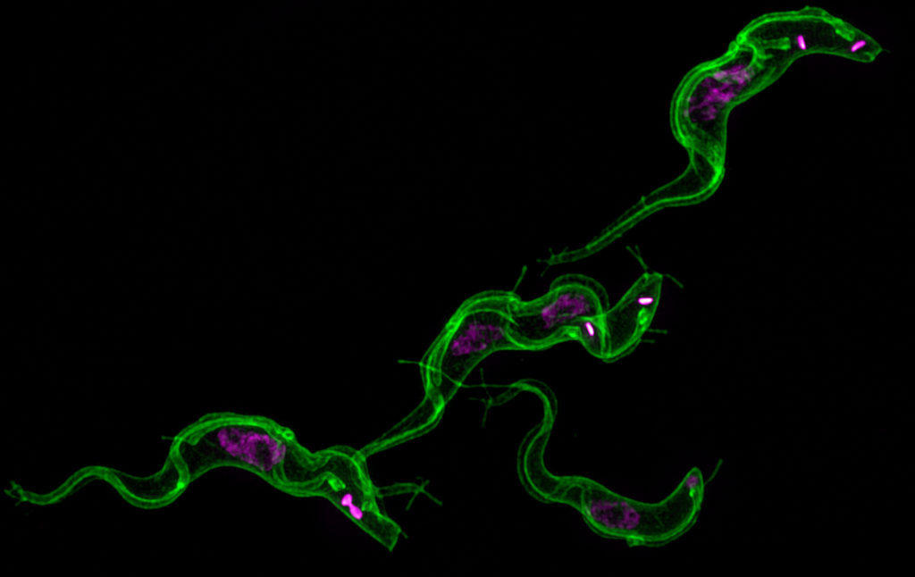
Trypanosoma brucei stained with mCLING
mCLING-staining of membrane (green) and DAPI staining of DNA (magenta) in trypanosomes in various life cycle stages. Flagella appear as tube-like structures along the length of the cell body. Nanotubes can be seen projecting outward from the periphery of the cells.

When Ph.D. trainee Justin Wiedeman started investigating the role of protein kinase TbCK1.2, an enzyme found near the flagellum of Trypanosoma brucei, he quickly ran into a problem common to parasitologists. He needed a better tool for visualizing the membranes of this parasite. Since none of the membrane probes on the market quite did the job, he looked at how he could modify one for his purpose. He found a successful candidate in Synaptic Systems’ mCLING.

What is Trypanosoma brucei?

Trypanosoma brucei is a single cell parasite that causes Human African Trypanosomiasis (HAT), which is also known as African sleeping sickness. HAT occurs in 36 sub-Saharan countries where tsetse flies transmit the parasite to people and livestock. In cattle, the disease is known as nagana. Tsetse fly control efforts have drastically reduced the number of cases. According to the World Health Organization, in 2015, there were around 2,800 cases. However, a person can be infected for months or even years without symptoms. By the time symptoms become evident, the person is in the advanced stages of the disease and their central nervous system is impaired.

New tools are needed to study trypanosomes

There is still much to be learned about the parasite that could lead to better detection and more effective treatment. A major obstacle to the study of this tiny organism is the lack of tools and technology. Kojo Mensa-Wilmot’s research group in the Center for Tropical and Emerging Global Diseases at The University of Georgia has been instrumental in developing techniques and tools to increase the research community’s understanding of T. brucei. Now, Wiedeman has added a new tool to the trypanosome biology toolbox – a general method of outlining trypanosomes in fluorescence microscopy experiments.

“We are the first group to solve this general problem in super-resolution microscopy of T. brucei,” said Wiedeman. “mCLING is a highly versatile tool for studying trypanosome biology – it can be used with live or fixed trypanosomes.”

Fluorescent microscopy has been a leading method of studying T. brucei; however, there are limitations to this technology. Super-resolution microscopy offers great advantages over standard fluorescence microscopy. By employing several techniques to increase resolution, it allows for the observation of objects smaller than what can be seen with visible light.  Yet, it is not without its own limitations, most notably the inability to determine the periphery of cells. Without knowing the outer edges of the parasite, orientation of organelles and other structures within the cell is difficult.

“For Trypanosoma brucei, most of the membrane probes available do not work well in fixed trypanosomes,” said Wiedeman. “Researchers have been forced to use crude methods to outline trypanosomes in fluorescence microscopy.”

These “crude methods” include superimposing a transmitted light image or hand-drawing the outline. However, this workaround only allows for a two-dimensional study of the cell. Therefore, Wiedeman turned to a dye called mCLING that has been developed to track the membranes of neurons using super-resolution microscopy to see if he could adapt the technology to T. brucei membranes.

mCLING allows for the visualization of T. brucei membranes

“mCLING labels the flagellum and plasma membrane vividly, sometimes providing details of cell structure that rivals images obtained with scanning electron microscopy,” said Wiedeman.

Using a combination of standard-resolution and super-resolution fluorescence microscopy, he was able to confirm mCLING labels the plasma and flagellar membranes of T. brucei. Furthermore, using the Zeiss ELYRA S1 super-resolution microscopy in the Biomedical Microscopy Core, mCLING allowed for a 3D reconstruction of the parasite. This is the first time such an image has been reported. Finally, using the new ImageStream X Mark II in the CTEGD Cytometry Shared Resource Laboratory, he discovered mCLING could be used to track endocytosis (the process of importing molecules into the cell) in real time.

Recognizing mCLING’s potential to inform other studies of trypanosome biology, Weideman optimized protocols for using it with immunofluorescence assays and thus making possible what had been impossible with the overlay technique – visualizing the location of organelles in the vertical dimension relative to the cell body.

“It is especially well-suited for studying flagellar membrane biogenesis as well as kinetically tracking uptake of the plasma membrane into vesicles inside trypanosomes,” said Wiedeman. Other laboratories have already implemented these protocols in their own research. Steve Hajduk’s group, also in the Center for Tropical and Emerging Global Diseases, is using mCLING to study nanotubes in T. brucei.

This tool will allow for the study of trypanosomes in finer detail than ever before and the Mensa-Wilmot Research Group anticipates unlocking previously unseen secrets in T. brucei.


The full published study is available online: Wiedeman J, Mensa-Wilmot K (2018). A fixable probe for visualizing flagella and plasma membranes of the African trypanosome. PLoS One 13(5):e0197541. https://doi.org/10.1371/journal.pone.0197541

Study reveals key cause of treatment failure in Chagas disease

Rick Tarleton
Photo by Peter Frey

Researchers at the University of Georgia have discovered that dormancy of the parasite Trypanosoma cruzi prevents effective drug treatment for Chagas disease, which kills more than 50,000 people each year in Central and South America and is a growing threat in the United States and Europe.

The disease infects an estimated 6 million to 7 million people, according to the World Health Organization, although some scientists estimate the number could be as high as 20 million. Chagas disease causing irreparable damage to the heart and digestive system, and effective prevention and treatment methods are virtually nonexistent.

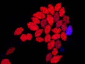
Proliferating Tdtomato expressing Trypanosoma cruzi amastigotes dilute the violet dye staining while non-replicating dormant parasite in the same host cell retains the violet signal.

In a new study published in eLife, Rick Tarleton and his research team at the Center for Tropical and Emerging Global Diseases sought to determine why drug treatments such as benzimidazole frequently fail.

“Benzimidazole has been shown to be particularly effective in reducing parasite infection,” said Tarleton, Regents’ Professor in the department of cellular biology.  “A single dose can eliminate nearly 90 percent of parasites within 48 hours, but we didn’t know why it didn’t kill 100 percent of the parasites.”

For the first time, they show that a small proportion of T. cruzi parasites halt replication within 24 hours of invading the host cell. These dormant parasites are resistant to extended drug treatment and can resume replication after treatment ends, thus re-establishing a growing infection.

The researchers don’t know why some of the parasites exhibit this behavior, but they are hopeful that future studies into this mechanism will shed more light on the way T. cruzi evades the host’s immune response.

“This isn’t drug resistance in the classical way we think of resistance,” said Tarleton. “The parasites aren’t dormant because of the presence of the drug.”

In fact, while treatment continued they saw some of the dormant parasites “wake up” and then become susceptible to the treatment. The team believes the key to effective treatment will be to catch the parasite as they resume replication, continuing medication until no parasites remain in the host.

“This discovery really offers a solution for current drugs to be used in a more effective way,” said Tarleton. “A longer, less concentrated dosing schedule could lead to a cure.”

T. cruzi lifecycle
Life cycle of Trypanosoma cruzi, the cause of Chagas disease (graphic by Lindsay Robinson


An online version of the study is available: https://elifesciences.org/articles/34039

An ancient bacterial protein complex in human malaria parasites is essential for parasite growth

Vasant Muralidharan and Anat Florentin

Several species of Plasmodium parasites cause malaria in humans and results in nearly 450,000 deaths annually. The deadliest of these species is Plasmodium falciparum. Unfortunately, it is also drug resistance to many of the currently available treatments. Vasant Muralidharan, assistant professor in the department of cellular biology, and his research group at The Center for Tropical and Emerging Global Diseases at The University of Georgia reported on an essential protein in hopes of identifying new drug targets.

Plasmodium parasites contain an organelle known as the apicoplast that evolved via the endosymbiosis of a red alga. The apicoplast produces several essential metabolites required for parasite growth and survival. Therefore, drugs that target the apicoplast are clinically effective. However, there is still not a lot known about this organelle. Understanding the function, structure, and biogenesis of the apicoplast provides a gold mine of antimalarial drug targets.

The role of Clp proteins in Plasmodium apicoplast

Clp (Caseinolytic Proteases) are conserved prokaryotic proteins that serve a wide variety of biological functions in bacteria, the evolutionary ancestors of the apicoplast. Several Clp proteins have been reported to localize in the apicoplast of the parasite but their biological functions were unknown.

The research team used different genetic tools to conditionally inhibit the function of various apicoplast-Clp proteins. “It is similar to understanding the role of a single card in holding up a house of cards by removing it from the structure,” said Muralidharan.

Their data show that the Clp chaperone PfClpC is essential for parasite viability and that its inhibition resulted in morphological defects, and loss of the apicoplast. They also revealed that the chaperone activity is required to stabilize a Clp Protease, PfClpP, suggesting that, similar to bacteria and plants chloroplasts, these two proteins form a proteolytic complex. These data may be relevant to the function of bacterial and plant Clp complexes. “Our findings shed light on the biological roles of the apicoplast Clp Proteins and their involvement in apicoplast replication,” said Dr. Anat Florentin, lead author on the study.

Significance of the findings

The role that bacterial Clp proteins play in cell division, stress response and ability to cause disease have placed them at the center of several drug discovery programs. The new understanding of Clp proteins in Plasmodium provides an avenue for drug development in malaria in which highly active antibacterial compounds can be repurposed as effective anti-malarial agents.


An online version of this study is available: A. Florentin,  D.W. Cobb, J.D. Fishburn, M. J. Cipriano, P.S. Kim, M.A. Fierro, B. Striepen, V. Muralidharan. 2017. PfClpC is an essential Clp chaperone required for plastid integrity and Clp protease stability in Plasmodium falciparum. Cell Reports 21, 1 – 11. http://dx.doi.org/10.1016/j.celrep.2017.10.081

A Double Hit Strategy May Provide Better Treatment for Toxoplasmosis

Zhu Hong Li
Zhu Hong Li is the lead author on the study.

Silvia Moreno and her research team at the University of Georgia’s Center for Tropical & Emerging Global Diseases and Department of Cellular Biology provided evidence that it is possible to develop a drug combination that acts synergistically by inhibiting host and parasite enzymes in a recently published article in Antimicrobial Agents and Chemotherapy.

What is toxoplasmosis?

Toxoplasmosis is caused by the pervasive intracellular Apicomplexan parasite Toxoplasma gondii. The parasite is found throughout the world and can infect humans and a number of animal species. In the U.S., people may contract it by consuming undercooked meats, especially pork, lamb, venison, or through contact with contaminated cat feces.

Human infections are usually asymptomatic but the parasite can persist in the form of tissue cysts. It has been estimated that 30–50% of the global population may be chronically infected with Toxoplasma. The immune system of a healthy individual can control the infection, but it can reactivate when there is immunosuppression due to organ transplant, cancer chemotherapy, or in people infected with HIV.

Toxoplasmosis is especially dangerous to the unborn fetus when the mother becomes infected during pregnancy as it can result in miscarriage or stillbirth. Surviving infants can suffer from visual, hearing, motor, cognitive, and other problems.

Some strains of T. gondii can cause severe ocular disease in people with a healthy immune system. Current drug therapies do not prevent disease progression that leads to blindness in ocular toxoplasmosis patients.

Toxoplasmosis represents a serious public health problem and no preventative or therapeutic vaccine is available for humans.


Need for better drug treatments for toxoplasmosis

Toxoplasma gondii
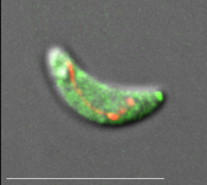
A Toxoplasma gondii parasite with cytosolic Green fluorescent protein and the mitochondrial red fluorescent marker.

Drugs presently used against toxoplasmosis do not eradicate chronic infection and as many as half of treated patients do not respond to the therapy. Additionally, a large number of people have an allergic reaction to the current treatment option. Furthermore, some the current drugs have recently become very expensive.

There is a need for safe and effective treatment.

Moreno and her team study the isoprenoid pathway to identify new drug targets. Isoprenoids are lipid compounds with many important functions. One particular step in this pathway has been identified as essential in T. gondii. A drug targeting this pathway could kill the parasite.

Drug combination may provide more effective and less expensive treatment

Moreno’s group proposes a double hit strategy of combining inhibitors of host and parasite pathways as a novel approach against toxoplasmosis. They have found a synergistic effect by combining new and potent sulfur-containing bisphosphonates, as well as other commercially available bisphosphonates, with several statins against a lethal infection of T. gondii using a virulence mouse model.

Bisphosphonates are widely used for the treatment of bone disorders. Previous studies by Moreno and her colleagues have shown that bisphosphonates inhibit the growth of a variety of protozoan parasites like T. gondii. There are a number of commercially available bisphosphonate drugs.

Statins are a class of drugs typically prescribed to lower cholesterol. They work by blocking a particular enzyme known as 3-hydroxy-methylglutaryl-coenzyme A reductase. As with the bisphosphonates, there are already a number of commercially available statins.

Bisphosphonates alone have been very effective when treating a lethal infection of T. gondii in mice. However, Moreno’s team found that combining bisphosphonates with the statin atorvastatin (Lipitor) was almost 3 times more effective under similar conditions of infection and treatment. Additionally, they found very low doses of both drugs could be used for treatment, which would significantly decrease the potential for adverse side effects.

This double hit strategy may be the key to effective treatment because the parasite not only makes its own isoprenoids, but it can also import them from the host. The ability to manipulate the host cell for its own benefit poses a challenge for drug development against toxoplasmosis. Therefore, inhibiting the host from producing this material along with inhibiting the parasite’s ability to create isoprenoids is an interesting and novel strategy for drug development.

Further studies for this novel therapeutic approach needed

This study demonstrates that early treatment is key to the cure of infection with a particular strain of T. gondii for acute infection. Since current treatments often fail to cure chronic infection Moreno and her group will next test this combination strategy in an established chronic infection mouse model.

Furthermore, Moreno predicts that this double-hit strategy of inhibiting both host and parasite pathways will work for other intracellular Apicomplexan parasites, such as the malaria-causing Plasmodium parasite. Additional studies will be needed to test this hypothesis.

An online version of this study is available: Li Z-H, Li C, Szajnman SH, Rodriguez JB, Moreno SNJ. 2017. Synergistic activity between statins and bisphosphonates against acute experimental toxoplasmosis. Antimicrob Agents Chemother 61:e02628-16. https://doi.org/10.1128/AAC.02628-16


The Cure for Malaria Could be in Your Backyard

Southern Magnolia

Malaria is a mosquito-borne disease that has a disproportionate effect in poor and underdeveloped countries without access to western medicine. According to the WHO’s World Malaria Report, there were 212 million new cases of malaria worldwide in 2015 and an estimated 429,000 deaths.

Malaria is caused by the Plasmodium parasite. Plasmodium falciparum is the deadliest of the species infecting humans, causing 50% of all malaria cases. Unfortunately, it has become resistant to current drug treatment.

Belen Cassera and her laboratory group at the University of Georgia have been identifying possible compounds for new antimalarial medication from natural plant sources.

Cassera Lab Group
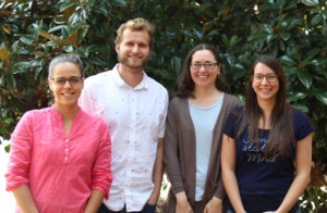
Cassera’s natural products team

History of anti-malarial drugs

Malaria has long been treated with plant-based medicine. Quinine, which comes from the bark of a cinchona tree, was first isolated as an antimalarial compound in the 1800s, though there is evidence that bark extracts have been used to treat malaria since the 1600s. The cinchona tree is native to Peru.

Quinine was the treatment of choice until the 1940s when other drugs, with fewer side effects, replaced it. One of those drugs was chloroquine, which was discovered in 1934. Following World War II, chloroquine became the preferred treatment for malaria and was prominent in mass drug administration programs of the 1950s. This wide-spread use, in part, led to chloroquine resistant strains of P. falciparum.

The rise of chloroquine-resistance led to the discovery of several potential synthetic alternatives. However, in 1972 Chinese scientists isolated artemisinin from Artemisia annua, commonly known as sweet wormwood. It is native to Asia but has been naturalized in several regions including North America. It has been the main treatment of malaria in south-east Asia. However, in recent years artemisinin resistance has also emerged. (Source: History of Antimalarials)

Cassera in collaboration with David Kingston at Virginia Tech and Michael Goetz and Jason Clement from the Natural Product Discovery Institute (NPDI) has a grant from the National Center for Complementary and Integrative Health (R01 AT008088) to study plants in the NPDI Repository to identify new antimalarial compounds.

Discovering new drugs from plants

In this project, they are concentrating on plants that have not been studied for their anti-malarial properties. Also, they are looking at plants that indigenous people have used to treat the various symptoms associated with malaria.

So far over 28,000 extracts have been screened and the team has identified over 100 compounds with anti-malarial activity.

In recently published findings, the group has reported the discovery of anti-malarial compounds in Malleastrum sp., Crinum firmifolium, and Magnolia grandiflora. The first two are plants found in Madagascar, but the last one is better known as the southern magnolia and can be found in backyards throughout the southeastern United States.

From the southern magnolia extracts, the Cassera and Kingston labs identified two new compounds with anti-malarial activity. It was also discovered that it contained six compounds that have been identified in other plants as possible malaria drug compounds. An online version of the study is available:  https://doi.org/10.1002/cbdv.201700209

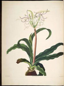
Crinumerubescens
By Bury, Edward; Havell, Robert [Public domain], via Wikimedia Commons
Extracts from Crinum species, which are in the amaryllis family, have been used traditionally to treat ailments including fever, pain management, swelling, sores and wounds, cancer, and malaria. Following success in isolating new anti-malarial compounds from an extract of Crinum erubescens L. F. ex Aiton, they turned to C. firmifolium, which was already in their International Cooperative Biodiversity Group collection. Extracts yielded 4 known compounds and three new compounds with possible anti-malarial activity. It was observed that the potency of several of the compounds against the drug-resistant strain of P. falciparum was approximately the same as their potency against the drug-sensitive strain. An online version of the study is available: https://doi.org/10.1016/j.bmc.2017.06.017

An extract of the wood from a species of Malleastrum in the mahogany family was found to have moderate antimalarial activity against a drug-resistant strain of P. falciparum. The genus Malleastrum (Baill.) J.-F. Leroy is endemic to Madagascar and comprises 20 currently accepted species. However, there appear to be at least four previously unidentified species. The plant material in this study is almost certainly from one of the species that is still waiting to be named and described. An online version of the study is available: https://doi.org/10.1002/cbdv.201700331

Next steps in the drug discovery process

“We have identified some really promising compounds,” said Belen Cassera. “A few could be ready for pre-clinical studies in a few years.”

In addition to testing for anti-malarial activity, the Cassera lab is also looking at the mechanism of action – how the compound works. This is an important step in drug discovery; because once it is understood how the compound works a synthetic analog could be synthesized and manufactured at a cheaper cost and in a safer form.

Each compound they have identified has several molecules associated with it. In their current state, some of these compounds have too high of a toxicity to be considered for potential drug treatment. Therefore, it is important to strip the compound down to only those molecules that have potent antimalarial responses and hopefully they can remove the molecules associated with toxicity.  Once this has been accomplished, then matching synthetic molecules can be created in the lab and scaled up for mass production.

Fun factBeing able to create these synthetic molecules is a necessary step in the drug discovery process. Natural material can be costly to collect or not available in abundance. While the southern magnolia seems to be abundant in the yards of Georgia, its natural range only stretches from coastal North Carolina south to central Florida, and then west to eastern Texas and Oklahoma. In addition, due to environmental differences, many times compounds isolated from a plant from one part of the world cannot be found in the same plant grown in another which reinforces the need to focus on active compounds that can be resynthesized in the lab.

With the appearance of drug-resistant strains of the malaria parasite to all current medications, it is imperative new treatments be discovered. Since plant-based and traditional medicine have yielded a number of drugs historically it is likely that the next treatment option will again come from a plant source. There are countless numbers of plants that have yet to be studied for their anti-malarial uses. Belen Cassera and her team just might find the cure for malaria in your backyard.

UGA researchers report milestone in global fight against a major cause of diarrheal disease

Sumiti Vinayak and Boris Striepen
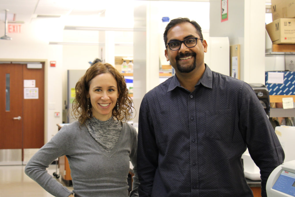
Assistant research scientist Sumiti Vinayak, left, and Distinguished Research Professor Boris Striepen work together in Striepen’s lab in the Coverdell Center for Biomedical and Health Sciences. Striepen and Vinayak are working together on vaccine and drug research for cryptosporidiosis, a disease caused by cryptosporidium, a microscopic parasite commonly spread through tainted drinking or recreational water, and it is a major cause of diarrheal disease and mortality in young children around the world. Credit: Andrew Davis Tucker, University of Georgia


Athens, Ga. – Infectious disease scientists from research institutions including the University of Georgia have reported the discovery and early validation of a drug that shows promise for treating cryptosporidiosis, a diarrheal disease that is a major cause of child mortality and for which there is no vaccine or effective treatment.

“Cryptosporidiosis is largely a disease of poverty,” said Boris Striepen, Distinguished Research Professor of Cellular Biology in UGA’s Franklin College of Arts and Sciences and a member of the Center for Tropical and Emerging Global Diseases. “Globally, it primarily affects infants in developing countries, but there are patients in the U.S.-those with weakened immune systems, such as HIV/AIDS or transplant patients-that would benefit greatly from new therapeutics.”

Striepen began studying crypto, as researchers often call the parasite that causes cryptosporidiosis, more than a decade ago. Now he and Sumiti Vinayak, assistant research scientist at UGA’s Center for Tropical and Emerging Global Diseases, along with scientists at Novartis and Washington State University, have reported the discovery of KDU731, a potent inhibitor of cryptosporidium, in the journal Nature.

Identifying KDU731 as a potential drug for the treatment of cryptosporidiosis began with the screening of a selection of 6,200 compounds that showed strong activity against the related malaria parasite. The Novartis team led by Ujjini H. Manjunatha and Thierry T. Diagana identified compounds with activity against crypto and found KDU731 particularly promising based on preclinical data.

Using a new mouse model, UGA’s Striepen and Vinayak showed that oral treatment with the drug dramatically reduced intestinal infection of immunocompromised mice. Additional research, led by Jennifer A. Zambriski at Washington State University, showed that treatment with KDU731 also leads to rapid resolution of diarrhea and dehydration in neonatal calves, a clinical model of cryptosporidiosis that closely resembles human infection.

Crypto is most commonly spread through tainted drinking or recreational water. When a person drinks contaminated water, parasites emerge from spores and invade the cells that line the small intestine, causing severe diarrhea that can last for up to three weeks.

In 1993, more than 400,000 people living in the Milwaukee, Wisconsin, area were infected and became ill when one of the city’s water treatment systems malfunctioned. More than 100 people, mostly AIDS patients, died during the outbreak.

Outbreaks have also been linked to swimming pools and water parks. Crypto is the most common cause of diarrheal illness and outbreaks linked to recreational water because it is not easily killed by chlorine and can survive up to 10 days in properly treated water.

The Centers for Disease Control and Prevention reported at least 32 outbreaks in U.S. facilities during 2016-twice as many as in 2014, according to preliminary data in the agency’s May 18 Morbidity and Mortality Weekly Report.

Recent global studies have shown crypto to be one of the most important causes of life-threatening diarrhea in infants and toddlers, especially in areas that lack access to clean water. There is no vaccine and only one drug, nitazoxanide, approved by the U.S. Food and Drug Administration, but it provides no benefit for those in gravest danger-malnourished infants and immunocompromised patients.

Crypto is notoriously difficult to work with in a laboratory setting, but Striepen has developed new genetic techniques that make it easier to detect and follow the parasite. One technique involves manipulating crypto so that it emits light and is easier to detect and measure. For this study, Striepen’s team engineered a new “reporter” parasite that is amenable to whole-animal imaging, allowing the researchers to non-invasively track and record dissipation of the infection during treatment.

Striepen’s genetically modified organisms have been made available to researchers across the world in the hope that more scientists will be drawn to studying crypto.

“This is an important problem,” he said. “No one institution can solve it alone. It needs significant investment, and it needs a lot of people with good ideas.”

“The discovery of this compound represents an important step toward urgently needed treatment for gravely ill children around the world,” said Thierry Diagana, head of the Novartis Institutes for Tropical Diseases.

An online version of the study is available at http://dx.doi.org/10.1038/nature22337.


Writer: Allyson Mann
Contact:Boris Striepen Sumiti Vinayak