Generic selectors
Exact matches only
Search in title
Search in content
Post Type Selectors

Category: publications

Multiple endocrine factors regulate nutrient mobilization and storage in Aedes aegypti during a gonadotrophic cycle

Anautogenous mosquitoes must blood feed on a vertebrate host to produce eggs. Each gonadotrophic cycle is subdivided into a sugar-feeding previtellogenic phase that produces primary follicles and a blood meal-activated vitellogenic phase in which large numbers of eggs synchronously mature and are laid. Multiple endocrine factors including juvenile hormone (JH), insulin-like peptides (ILPs), ovary ecdysteroidogenic hormone (OEH) and 20-hydroxyecdysone (20E) coordinate each gonadotrophic cycle. Egg formation also requires nutrients from feeding that are stored in the fat body. Regulation of egg formation is best understood in Aedes aegypti but the role different endocrine factors play in regulating nutrient mobilization and storage remains unclear. In this study, we report that adult female Ae. aegypti maintained triacylglycerol (TAG) stores during the previtellogenic phase of the first gonadotrophic cycle while glycogen stores declined. In contrast, TAG and glycogen stores were rapidly mobilized during the vitellogenic phase and then replenishment. Several genes encoding enzymes with functions in TAG and glycogen metabolism were differentially expressed in the fat body, which suggested regulation was mediated in part at the transcriptional level. Gain of function assays indicated that stored nutrients were primarily mobilized by adipokinetic hormone (AKH) while juvenoids and OEH regulated replenishment. ILP3 further showed evidence of negatively regulating certain lipolytic enzymes. Loss of function assays further indicated AKH depends on the AKH receptor (AKHR) for function. Altogether, our results indicate that the opposing activities of different hormones regulate nutrient stores during a gonadotrophic cycle in Ae. aegypti. This article is protected by copyright. All rights reserved.

Xiaoyi Dou, Kangkang Chen, Mark R Brown, Michael R Strand. Insect Sci. 2022 Sep 2. doi: 10.1111/1744-7917.13110.

Single-cell RNA profiling of Plasmodium vivax-infected hepatocytes reveals parasite- and host- specific transcriptomic signatures and therapeutic targets

The resilience of Plasmodium vivax, the most widely-distributed malaria-causing parasite in humans, is attributed to its ability to produce dormant liver forms known as hypnozoites, which can activate weeks, months, or even years after an initial mosquito bite. The factors underlying hypnozoite formation and activation are poorly understood, as is the parasite’s influence on the host hepatocyte. Here, we shed light on transcriptome-wide signatures of both the parasite and the infected host cell by sequencing over 1,000 P. vivax-infected hepatocytes at single-cell resolution. We distinguish between replicating schizonts and hypnozoites at the transcriptional level, identifying key differences in transcripts encoding for RNA-binding proteins associated with cell fate. In infected hepatocytes, we show that genes associated with energy metabolism and antioxidant stress response are upregulated, and those involved in the host immune response downregulated, suggesting both schizonts and hypnozoites alter the host intracellular environment. The transcriptional markers in schizonts, hypnozoites, and infected hepatocytes revealed here pinpoint potential factors underlying dormancy and can inform therapeutic targets against P. vivax liver-stage infection.

Anthony A Ruberto, Steven P Maher, Amélie Vantaux, Chester J Joyner, Caitlin Bourke, Balu Balan, Aaron Jex, Ivo Mueller, Benoit Witkowski, Dennis E Kyle. Front Cell Infect Microbiol. 2022 Aug 25;12:986314. doi: 10.3389/fcimb.2022.986314. eCollection 2022.

Temporal and thermal profiling of the Toxoplasma proteome implicates parasite Protein Phosphatase 1 in the regulation of Ca 2+-responsive pathways

Apicomplexan parasites cause persistent mortality and morbidity worldwide through diseases including malaria, toxoplasmosis, and cryptosporidiosis. Ca2+ signaling pathways have been repurposed in these eukaryotic pathogens to regulate parasite-specific cellular processes governing the replicative and lytic phases of the infectious cycle, as well as the transition between them. Despite the presence of conserved Ca2+-responsive proteins, little is known about how specific signaling elements interact to impact pathogenesis. We mapped the Ca2+-responsive proteome of the model apicomplexan T. gondii via time-resolved phosphoproteomics and thermal proteome profiling. The waves of phosphoregulation following PKG activation and stimulated Ca2+ release corroborate known physiological changes but identify specific proteins operating in these pathways. Thermal profiling of parasite extracts identified many expected Ca2+-responsive proteins, such as parasite Ca2+-dependent protein kinases. Our approach also identified numerous Ca2+-responsive proteins that are not predicted to bind Ca2+, yet are critical components of the parasite signaling network. We characterized protein phosphatase 1 (PP1) as a Ca2+-responsive enzyme that relocalized to the parasite apex upon Ca2+ store release. Conditional depletion of PP1 revealed that the phosphatase regulates Ca2+ uptake to promote parasite motility. PP1 may thus be partly responsible for Ca2+-regulated serine/threonine phosphatase activity in apicomplexan parasites.

Alice L Herneisen, Zhu-Hong Li, Alex W Chan, Silvia N J Moreno, Sebastian Lourido. Elife. 2022 Aug 17;11:e80336. doi: 10.7554/eLife.80336.

Parasite reliance on its host gut microbiota for nutrition and survival

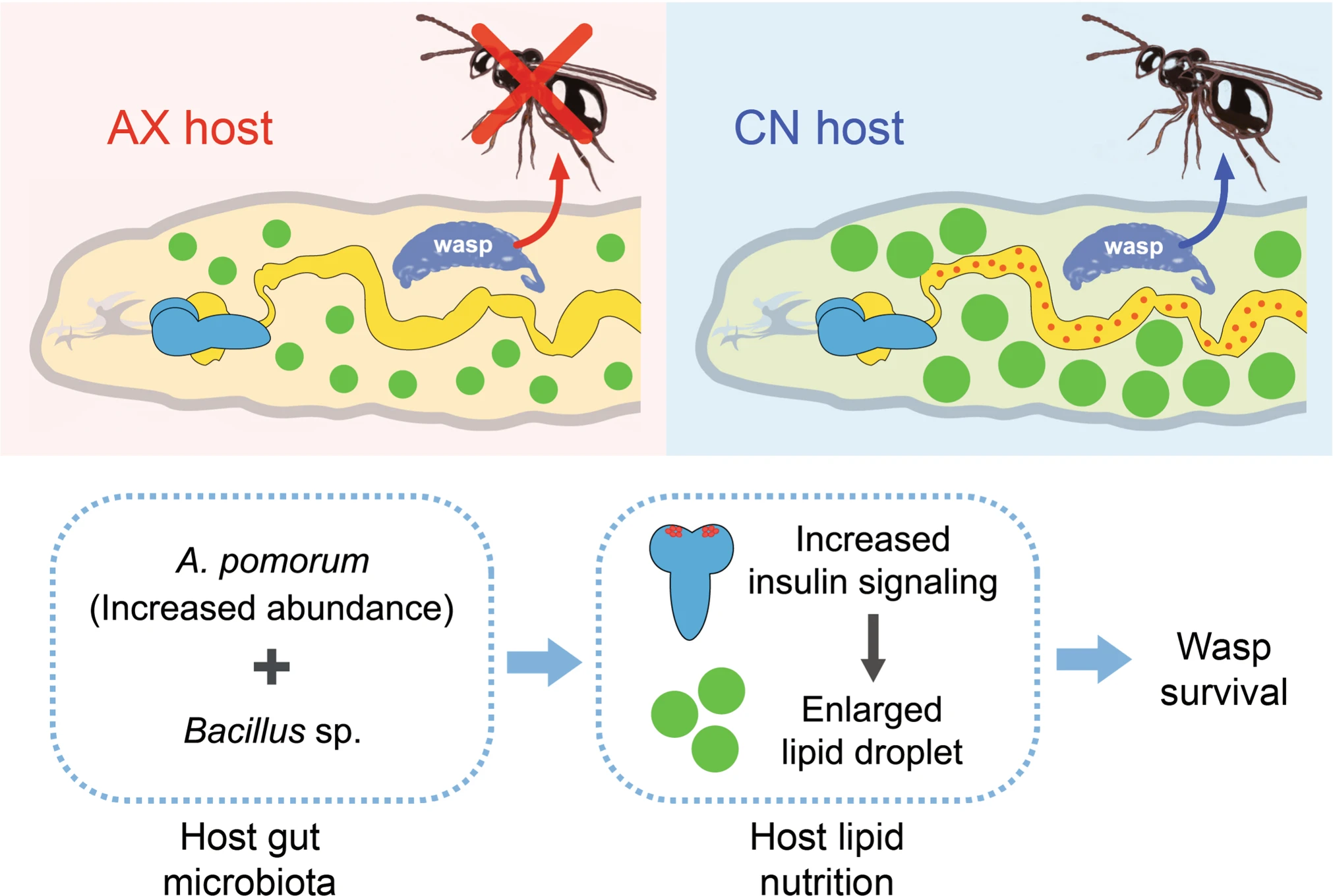
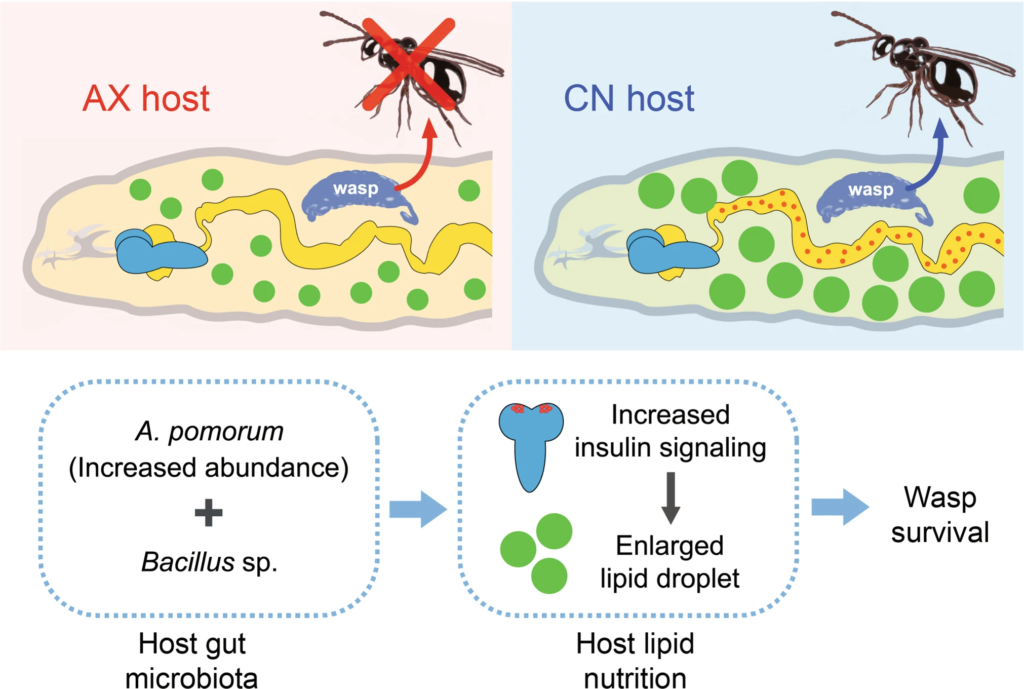
The proposed model of how host gut microbiota promotes parasite survival. (Figure 6)

Studying the microbial symbionts of eukaryotic hosts has revealed a range of interactions that benefit host biology. Most eukaryotes are also infected by parasites that adversely affect host biology for their own benefit. However, it is largely unclear whether the ability of parasites to develop in hosts also depends on host-associated symbionts, e.g., the gut microbiota. Here, we studied the parasitic wasp Leptopilina boulardi (Lb) and its host Drosophila melanogaster. Results showed that Lb successfully develops in conventional hosts (CN) with a gut microbiota but fails to develop in axenic hosts (AX) without a gut microbiota. We determined that developing Lb larvae consume fat body cells that store lipids. We also determined that much larger amounts of lipid accumulate in fat body cells of parasitized CN hosts than parasitized AX hosts. CN hosts parasitized by Lb exhibited large increases in the abundance of the bacterium Acetobacter pomorum in the gut, but did not affect the abundance of Lactobacillus fructivorans which is another common member of the host gut microbiota. However, AX hosts inoculated with A. pomorum and/or L. fructivorans did not rescue development of Lb. In contrast, AX larvae inoculated with A. pomorum plus other identified gut community members including a Bacillus sp. substantially rescued Lb development. Rescue was further associated with increased lipid accumulation in host fat body cells. Insulin-like peptides increased in brain neurosecretory cells of parasitized CN larvae. Lipid accumulation in the fat body of CN hosts was further associated with reduced Bmm lipase activity mediated by insulin/insulin-like growth factor signaling (IIS). Altogether, our results identify a previously unknown role for the gut microbiota in defining host permissiveness for a parasite. Our findings also identify a new paradigm for parasite manipulation of host metabolism that depends on insulin signaling and the gut microbiota.

Sicong Zhou, Yueqi Lu, Jiani Chen, Zhongqiu Pan, Lan Pang, Ying Wang, Qichao Zhang, Michael R Strand, Xue-Xin Chen, Jianhua Huang. ISME J. 2022 Aug 8. doi: 10.1038/s41396-022-01301-z.

Onchocerciasis: Target product profiles of in vitro diagnostics to support onchocerciasis elimination mapping and mass drug administration stopping decisions

In June 2021, the World Health Organization (WHO), recognizing the need for new diagnostics to support the control and elimination of onchocerciasis, published the target product profiles (TPPs) of new tests that would support the two most immediate needs: (a) mapping onchocerciasis in areas of low prevalence and (b) deciding when to stop mass drug administration programs. In both instances, the test should ideally detect an antigen specific for live, adult O. volvulus female worms. The preferred format is a field-deployable rapid test. For mapping, the test needs to be ≥ 60% sensitive and ≥ 99.8% specific, while to support stopping decisions, the test must be ≥ 89% sensitive and ≥ 99.8% specific. The requirement for extremely high specificity is dictated by the need to detect with sufficient statistical confidence the low seroprevalence threshold set by WHO. Surveys designed to detect a 1-2% prevalence of a given biomarker, as is the case here, cannot tolerate more than 0.2% of false-positives. Otherwise, the background noise would drown out the signal. It is recognized that reaching and demonstrating such a stringent specificity criterion will be challenging, but test developers can expect to be assisted by national governments and implementing partners for adequately powered field validation.

Marco A. Biamonte ,Paul T. Cantey,Yaya I. Coulibaly,Katherine M. Gass,Louise C. Hamill,Christopher Hanna,Patrick J. Lammie,Joseph Kamgno,Thomas B. Nutman,David W. Oguttu,Dieudonné P. Sankara,Wilma A. Stolk,Thomas R. Unnasch. PLoS Negl Trop Dis. 2022 Aug 3;16(8):e0010682. doi: 10.1371/journal.pntd.0010682.

Oxygen-dependent regulation of E3(SCF)ubiquitin ligases and a Skp1-associated JmjD6 homolog in development of the social amoeba Dictyostelium

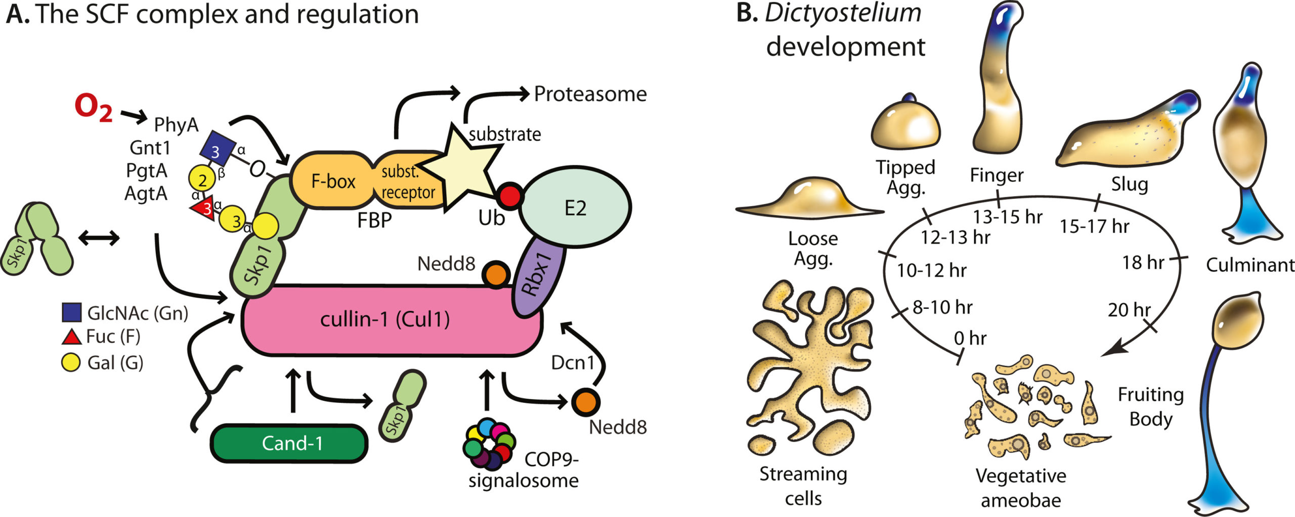
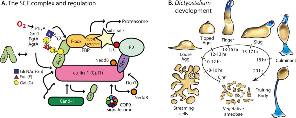
E3-SCF (Skp1/cullin-1/F-box protein) polyubiquitin ligases activate the proteasomal degradation of over a thousand proteins, but the evolutionary diversification of the F-box protein (FBP) family of substrate receptor subunits has challenged their elucidation in protists. Here we expand the FBP candidate list in the social amoeba Dictyostelium and show that the Skp1 interactome is highly remodeled as cells transition from solitary growth to multicellular development. Importantly, a subset of candidate FBPs was less represented when the posttranslational hydroxylation and glycosylation of Skp1 was abrogated by deletion of the O2 -sensing Skp1 prolyl hydroxylase PhyA. A role for this Skp1 modification for SCF activity was indicated by partial rescue of development, which normally depends on high O2 and PhyA, of phyA -knockout cells by proteasomal inhibitors. Further examination of two FBPs, FbxwD and the Jumonji C protein JcdI, suggested that Skp1 was substituted by other factors in phyA-knockout cells. Although a double-knockout of jcdI and its paralog jcdH did not affect development, overexpression of JcdI increased its sensitivity to O2. JcdI, a non-heme dioxygenase shown to have physiological O2-dependence, is conserved across protists with its F-box and other domains, and related to the human oncogene JmjD6. Sensitization of JcdI-overexpression cells to O2 depended on its dioxygenase activity and other domains, but not its F-box, which may however be the mediator of its reduced levels in wild-type relative to Skp1 modification mutant cells. The findings suggest that activation of JcdI by O2 is tempered by homeostatic down-regulation via PhyA and association with Skp1.

Andrew W Boland, Elisabet Gas-Pascual, Braxton L Nottingham, Hanke van der Wel, M Osman Sheikh, Christopher M Schafer, Christopher M West. J Biol Chem. 2022 Aug 3;102305. doi: 10.1016/j.jbc.2022.102305.

Direct type I interferon signaling in hepatocytes controls malaria

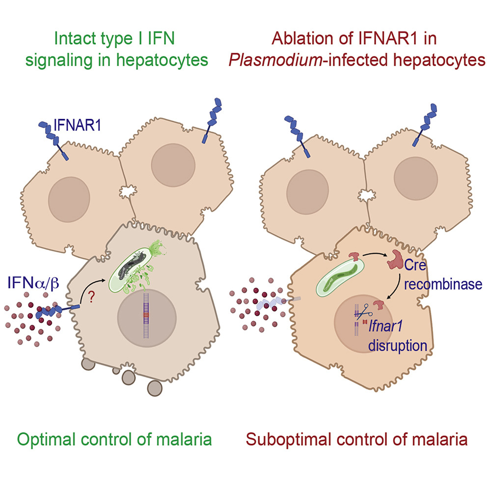
Malaria is a devastating disease impacting over half of the world’s population. Plasmodium parasites that cause malaria undergo obligatory development and replication in hepatocytes before infecting red blood cells and initiating clinical disease. While type I interferons (IFNs) are known to facilitate innate immune control to Plasmodium in the liver, how they do so has remained unresolved, precluding the manipulation of such responses to combat malaria. Utilizing transcriptomics, infection studies, and a transgenic Plasmodium strain that exports and traffics Cre recombinase, we show that direct type I IFN signaling in Plasmodium-infected hepatocytes is necessary to control malaria. We also show that the majority of infected hepatocytes naturally eliminate Plasmodium infection, revealing the potential existence of anti-malarial cell-autonomous immune responses in such hepatocytes. These discoveries challenge the existing paradigms in Plasmodium immunobiology and are expected to inspire anti-malarial drugs and vaccine strategies.

Camila Marques-da-Silva, Kristen Peissig, Michael P Walker, Justine Shiau, Carson Bowers, Dennis E Kyle, Rahul Vijay, Scott E Lindner, Samarchith P Kurup. Cell Rep. 2022 Jul 19;40(3):111098. doi: 10.1016/j.celrep.2022.111098.

A Krüppel-like factor is required for development and regeneration of germline and yolk cells from somatic stem cells in planarians

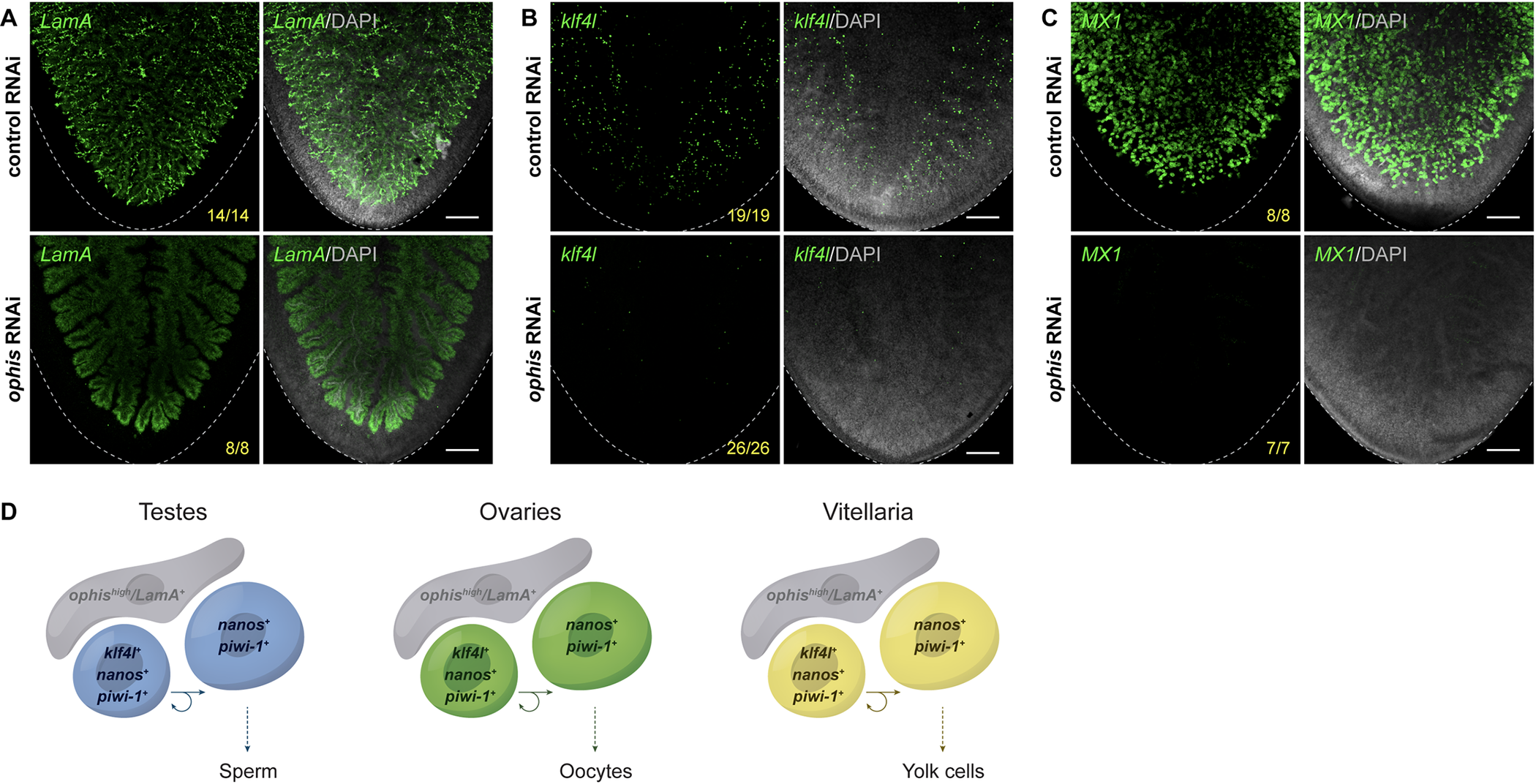
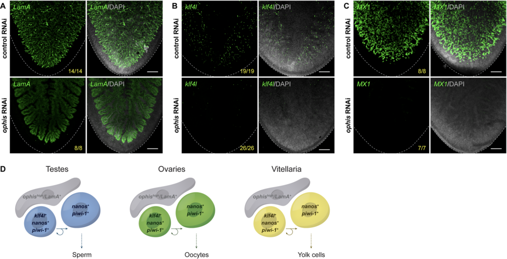
Fig 8. Germ cell niche factor ophis is required to sustain yolk cell production/vitellogenesis.
(A–C) Maximum intensity projections of confocal sections showing FISH of LamA (A), klf4l (B), and MX1 (C) (green) in the ventral posterior region of sexually mature control versus ophis RNAi animals. Dashed line denotes planarian boundary. N = 3 to 5 experiments, n = 7 to 26 planarians. (A) ophis RNAi results in a dramatic loss of the LamA+ cells throughout the vitellaria. Note that LamA expression is only visible in the branched gut in ophis RNAi planarians. (B, C) ophis RNAi results in a reduction of klf4l+ yolk cell progenitors and MX1+ differentiated yolk cells. (A–C) Nuclei are counterstained with DAPI (gray). Scale bars, 200 μm. (D) Model depicting similarities shared between gonads (where gametogenesis occurs) and vitellaria (where yolk cell production occurs). klf4l+/nanos+/piwi-1+ presumptive GSCs in testes and ovaries divide and give rise to klf4l–/nanos+/piwi-1+ progeny. These germ cells are supported by ophis+ somatic gonadal niche cells. Vitellaria are comprised of klf4l+/nanos+/piwi-1+ “germ cell–like” yolk progenitors that are mitotically competent, sustain yolk cell production, and are supported by ophishigh support cells. FISH, fluorescent RNA in situ hybridization; klf4l, klf4-like; RNAi, RNA interference.

Abstract

Sexually reproducing animals segregate their germline from their soma. In addition to gamete-producing gonads, planarian and parasitic flatworm reproduction relies on yolk cell-generating accessory reproductive organs (vitellaria) supporting development of yolkless oocytes. Despite the importance of vitellaria for flatworm reproduction (and parasite transmission), little is known about this unique evolutionary innovation. Here, we examine reproductive system development in the planarian Schmidtea mediterranea, in which pluripotent stem cells generate both somatic and germ cell lineages. We show that a homolog of the pluripotency factor Klf4 is expressed in primordial germ cells (PGCs), presumptive germline stem cells (GSCs), and yolk cell progenitors. Knockdown of this klf4-like (klf4l) gene results in animals that fail to specify or maintain germ cells; surprisingly, they also fail to maintain yolk cells. We find that yolk cells display germ cell-like attributes and that vitellaria are structurally analogous to gonads. In addition to identifying a new proliferative cell population in planarians (yolk cell progenitors) and defining its niche, our work provides evidence supporting the hypothesis that flatworm germ cells and yolk cells share a common evolutionary origin.

Melanie Issigonis, Akshada B Redkar, Tania Rozario, Umair W Khan, Rosa Mejia-Sanchez, Sylvain W Lapan, Peter W Reddien, Phillip A Newmark. PLoS Biol. 2022 Jul 15;20(7):e3001472. doi: 10.1371/journal.pbio.3001472.

Quantitative 3D Imaging of Trypanosoma cruzi-Infected Cells, Dormant Amastigotes, and T Cells in Intact Clarified Organs

Chagas disease is a neglected pathology that affects millions of people worldwide, mainly in Latin America. The Chagas disease agent, Trypanosoma cruzi (T. cruzi), is an obligate intracellular parasite with a diverse biology that infects several mammalian species, including humans, causing cardiac and digestive pathologies. Reliable detection of T. cruzi in vivo infections has long been needed to understand Chagas disease’s complex biology and accurately evaluate the outcome of treatment regimens. The current protocol demonstrates an integrated pipeline for automated quantification of T. cruzi-infected cells in 3D-reconstructed, cleared organs. Light-sheet fluorescent microscopy allows for accurately visualizing and quantifying of actively proliferating and dormant T. cruzi parasites and immune effector cells in whole organs or tissues. Also, the CUBIC-HistoVision pipeline to obtain uniform labeling of cleared organs with antibodies and nuclear stains was successfully adopted. Tissue clearing coupled with 3D immunostaining provides an unbiased approach to comprehensively evaluate drug treatment protocols, improve the understanding of the cellular organization of T. cruzi-infected tissues, and is expected to advance discoveries related to anti-T. cruzi immune responses, tissue damage, and repair in Chagas disease.

Fernando Sanchez-Valdez, Ángel M Padilla, Juan M Bustamante, Caleb W D Hawkins, Rick L Tarleton. J Vis Exp. 2022 Jun 23;(184). doi: 10.3791/63919.

Plasmodium knowlesi Cytoadhesion Involves SICA Variant Proteins

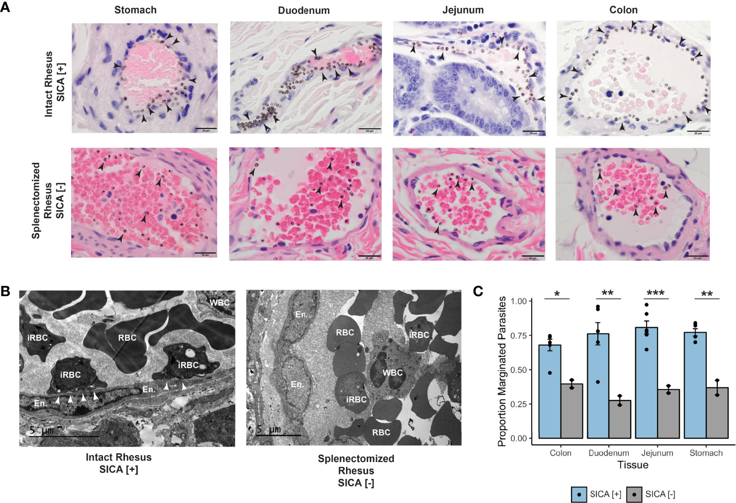
Plasmodium knowlesi poses a health threat throughout Southeast Asian communities and currently causes most cases of malaria in Malaysia. This zoonotic parasite species has been studied in Macaca mulatta (rhesus monkeys) as a model for severe malarial infections, chronicity, and antigenic variation. The phenomenon of Plasmodium antigenic variation was first recognized during rhesus monkey infections. Plasmodium-encoded variant proteins were first discovered in this species and found to be expressed at the surface of infected erythrocytes, and then named the Schizont-Infected Cell Agglutination (SICA) antigens. SICA expression was shown to be spleen dependent, as SICA expression is lost after P. knowlesi is passaged in splenectomized rhesus. Here we present data from longitudinal P. knowlesi infections in rhesus with the most comprehensive analysis to date of clinical parameters and infected red blood cell sequestration in the vasculature of tissues from 22 organs. Based on the histopathological analysis of 22 tissue types from 11 rhesus monkeys, we show a comparative distribution of parasitized erythrocytes and the degree of margination of the infected erythrocytes with the endothelium. Interestingly, there was a significantly higher burden of parasites in the gastrointestinal tissues, and extensive margination of the parasites along the endothelium, which may help explain gastrointestinal symptoms frequently reported by patients with P. knowlesi malarial infections. Moreover, this margination was not observed in splenectomized rhesus that were infected with parasites not expressing the SICA proteins. This work provides data that directly supports the view that a subpopulation of P. knowlesi parasites cytoadheres and sequesters, likely via SICA variant antigens acting as ligands. This process is akin to the cytoadhesive function of the related variant antigen proteins, namely Erythrocyte Membrane Protein-1, expressed by Plasmodium falciparum.

Mariko S Peterson, Chester J Joyner, Stacey A Lapp, Jessica A Brady, Jennifer S Wood, Monica Cabrera-Mora, Celia L Saney, Luis L Fonseca, Wayne T Cheng, Jianlin Jiang, Stephanie R Soderberg, Mustafa V Nural, Allison Hankus, Deepa Machiah, Ebru Karpuzoglu, Jeremy D DeBarry, Rabindra Tirouvanziam, Jessica C Kissinger, Alberto Moreno, Sanjeev Gumber, Eberhard O Voit, Juan B Gutierrez, Regina Joice Cordy, Mary R Galinski. Front Cell Infect Microbiol. 2022 Jun 23;12:888496. doi: 10.3389/fcimb.2022.888496. eCollection 2022.