Generic selectors
Exact matches only
Search in title
Search in content
Post Type Selectors

Author: Donna Huber

UGA researcher uncovers humans’ natural weapon against malaria

UGA’s Samarchith “Sam” Kurup, assistant professor of cellular biology, has been awarded a five-year National Institutes of Health grant to study the natural immune response to the Plasmodium parasite—which causes malaria—in liver cells. (photo credit: Lauren Corcino)

Samarchith “Sam” Kurup grew up in India, and he’s always been aware of the impact of malaria.

In 2020 there were an estimated 241 million cases of malaria worldwide and an estimated 627,000 deaths, according to a recently released World Health Organization Fact Sheet. Eighty percent of the malaria-related deaths in Africa are children under the age of 5. The relapsing nature of the disease leads to educational and employment loss that has long-term economic impacts for both the individual as well as society.

“Malaria is huge global problem,” said Kurup, a member of UGA’s Center for Tropical and Emerging Global Diseases. “Almost half of the world’s population is currently at risk of contracting malaria.”

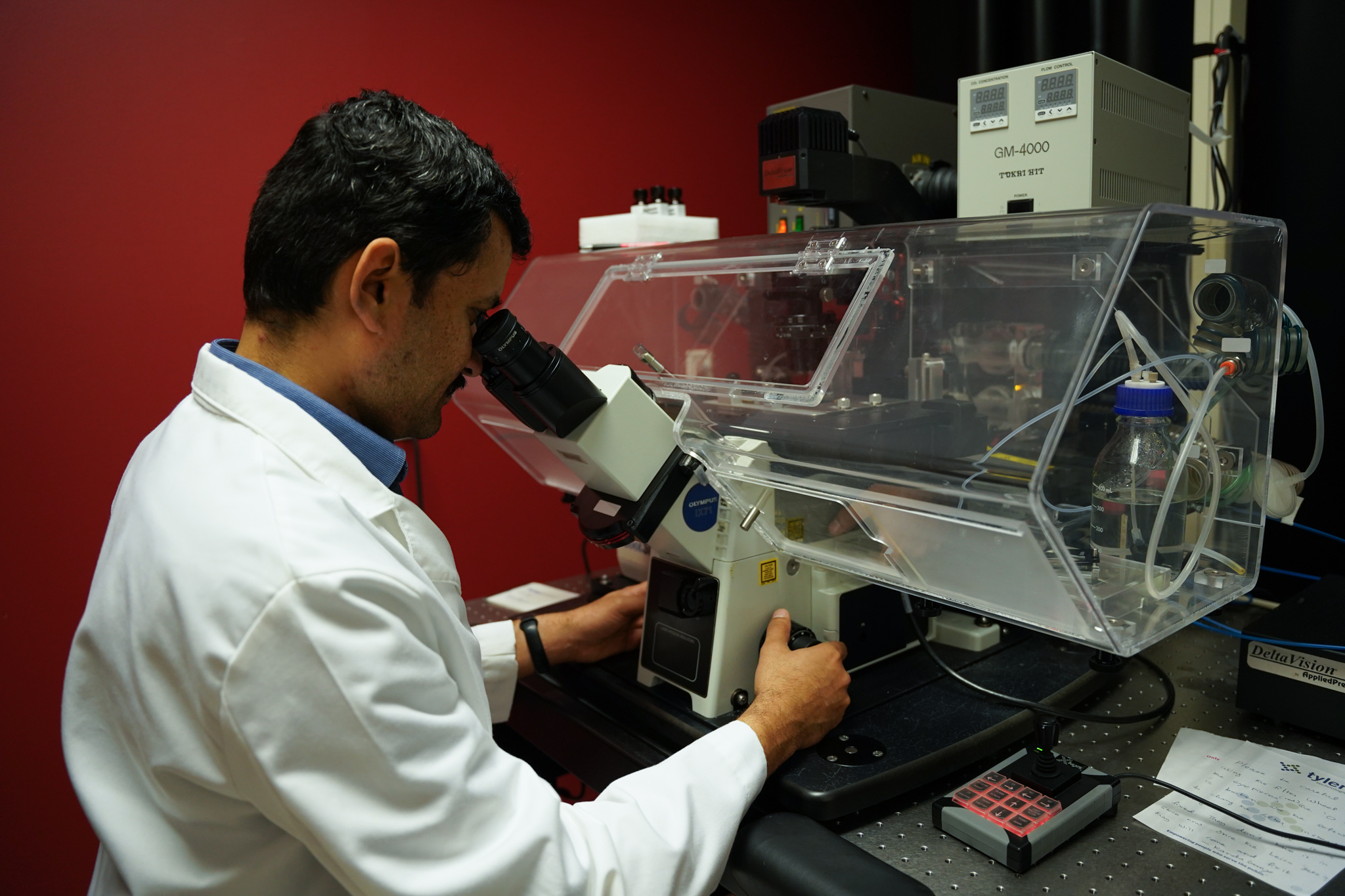
Kurup began his training in veterinary medicine in India, where he became hooked on parasitology, then continued his studies at UGA. While pursuing his Ph.D. he worked in Rick Tarleton’s lab, studying a parasitic disease that affects both animals and humans—his first introduction to human immunology. He continued his training in immunology as a postdoctoral researcher in John Harty’s lab at the University of Iowa.

Combining parasitology with immunology prepared him to tackle malaria.

Malaria is one of the most studied parasitic diseases, yet the Plasmodium parasite that causes it keeps evading attempts to treat the infection in humans. This is largely due to its complex life cycle and the ability of the parasite to evolve drug resistance. In addition to life stages that occur in the mosquito, which transmits the Plasmodium parasite to humans, there are two life stages in humans—a short phase of initial development in the liver, followed by an infection of the blood cells that causes clinical disease.

“A lot of research has been focused on the blood stage in humans, as this is when a person is symptomatic,” said Kurup, assistant professor of cellular biology in the Franklin College of Arts and Sciences. “But we now recognize that if we want to stop malaria, we need to stop it in its tracks in the liver before accessing the blood, and for that we need to understand the liver stage.”

Kurup, a member of UGA’s Center for Tropical and Emerging Global Diseases, trained in parasitology and immunology. He hopes that uncovering how the human immune system naturally fights malaria in the liver stage will lead to an effective malaria vaccine. (photo credit: Lauren Corcino)

Kurup has been awarded a five-year National Institutes of Health grant to study the natural immune response to the Plasmodium parasite in liver cells.

“The liver stage is short and can be difficult to study in the laboratory,” he said. “There are also practical and ethical limitations to studying the liver stage of malaria in humans. We are hoping to tease apart the basic principles of immune responses during this stage using the mouse model.”

Kurup’s preliminary studies have shown that a group of signaling proteins called type 1 interferons play a role in the destruction of Plasmodium parasites in the liver. His newly funded project will fill a gap in the malaria knowledge base by using a combination of in vitro study and in vivo experiments to determine the molecular processes that eliminate Plasmodium parasites in liver cells. His group recently developed a transgenic parasite line that can be used to genetically alter its host cell.

“This strain is a game changer for our line of research because we can now determine how our liver cells would naturally eliminate the parasite, and maybe why it sometimes fails,” he said.

In a study recently published in Cell Reports, Kurup and colleagues used the genetically altered parasite to inhibit signaling by type 1 interferons and showed that this protein has a direct role in the control of malaria. Their study also revealed that other natural immune mechanisms may be active in controlling malaria in liver cells. The project funded by the new grant will delve further into these mechanisms.

“In addition to taking us a step closer to the control and possible eradication of malaria, this project will expand our knowledge so that we can better reduce the burdens of this illness in our society,” he said.

Kurup is hopeful that uncovering how the human immune system naturally fights malaria in the liver stage will lead to an effective malaria vaccine.

“I really believe that bringing together our knowledge in parasitology and approaches in immunology is key to uncovering new information on this elusive life stage in malaria,” he said. “There is no better place to do this, considering the intellectual and material resources we have at our disposal at UGA and the CTEGD.”


This story was first published at https://research.uga.edu/news/uga-researcher-uncovers-humans-natural-weapon-against-malaria/

MICU1 and MICU2 potentiation of Ca2+ uptake by the mitochondrial Ca2+ uniporter of Trypanosoma cruzi and its inhibition by Mg2

Trypanosome MCU cimplex organization

The mitochondrial Ca2+ uptake, which is important to regulate bioenergetics, cell death and cytoplasmic Ca2+ signaling, is mediated via the calcium uniporter complex (MCUC). In animal cells the MCUC is regulated by the mitochondrial calcium uptake 1 and 2 dimer (MICU1/MICU2), which has been proposed to act as gatekeeper preventing mitochondrial Ca2+ overload at low cytosolic Ca2+ levels. In contrast to animal cells, knockout of either MICU1 or MICU2 in Trypanosoma cruzi, the etiologic agent of Chagas disease, did not allow Ca2+ uptake at low extramitochondrial Ca2+ concentrations ([Ca2+]ext) and it was though that in the absence of one MICU the other would replace its role. However, previous attempts to knockout both genes were unsuccessful. Here, we designed a strategy to generate TcMICU1/TcMICU2 double knockout cell lines using CRISPR/Cas9 genome editing. Ablation of both genes was confirmed by PCR and Southern blot analyses. The absence of both proteins did not allow Ca2+ uptake at low [Ca2+]ext, significantly decreased the mitochondrial Ca2+ uptake at different [Ca2+]ext, without dissipation of the mitochondrial membrane potential, and increased the [Ca2+]ext set point needed for Ca2+ uptake, as we have seen with TcMICU1-KO and TcMICU2-KO cells. Mg2+ was found to be a negative regulator of MCUC-mediated mitochondrial Ca2+ uptake at different [Ca2+]ext. Occlusion of the MCUC pore by Mg2+ could partially explain the lack of mitochondrial Ca2+ uptake at low [Ca2+]ext in TcMICU1/TcMICU2-KO cells. In addition, TcMICU1/TcMICU2-KO epimastigotes had a lower growth rate, while infective trypomastigotes have a reduced capacity to invade host cells and to replicate within them as amastigotes.

Mayara S Bertolini, Roberto Docampo. Cell Calcium. 2022 Sep 21;107:102654. doi: 10.1016/j.ceca.2022.102654.

The Domestic Dog as a Laboratory Host for Brugia malayi

Of the three nematodes responsible for lymphatic filariasis in humans, only Brugia malayi is actively maintained in research settings owing to its viability in small animal hosts, principal among which is the domestic cat. While the microfilaremic feline host is necessary for propagation of parasites on any significant scale, this system is plagued by a number of challenges not as pronounced in canine filarial models. For this reason, we investigated the capacity in which dogs may serve as competent laboratory hosts for B. malayi. We infected a total of 20 dogs by subcutaneous injection of 500 B. malayi third-stage larvae (L3) in either a single (n = 10) or repeated infection events (125 L3 per week for four weeks; n = 10). Within each group, half of the individuals were injected in the inguinal region and half in the dorsum of the hind paw. To track the course of microfilaremia in this host, blood samples were examined by microscopy biweekly for two years following infection. Additionally, to identify cellular responses with potential value as predictors of patency, we measured peripheral blood leukocyte counts for the first year of infection. A total of 10 of 20 dogs developed detectable microfilaremia. Peak microfilaria density varied but attained levels useful for parasite propagation (median = 1933 mL-1; range: 33-9950 mL-1). Nine of these dogs remained patent at 104 weeks. A two-way ANOVA revealed no significant differences between infection groups in lifetime microfilaria production (p = 0.42), nor did regression analysis reveal any likely predictive relationships to leukocyte values. The results of this study demonstrate the competence of the dog as a host for B. malayi and its potential to serve in the laboratory role currently provided by the cat, while also clarifying the potential for zoonosis in filariasis-endemic regions.

Christopher C Evans, Katelin E Greenway, Elyssa J Campbell, Michael T Dzimianski, Abdelmoneim Mansour, John W McCall, Andrew R Moorhead. Pathogens. 2022 Sep 21;11(10):1073. doi: 10.3390/pathogens11101073.

The Heptaprenyl Diphosphate Synthase (Coq1) Is the Target of a Lipophilic Bisphosphonate That Protects Mice against Toxoplasma gondii Infection

Prenyldiphosphate synthases catalyze the reaction of allylic diphosphates with one or more isopentenyl diphosphate molecules to form compounds such as farnesyl diphosphate, used in, e.g., sterol biosynthesis and protein prenylation, as well as longer “polyprenyl” diphosphates, used in ubiquinone and menaquinone biosynthesis. Quinones play an essential role in electron transport and are associated with the inner mitochondrial membrane due to the presence of the polyprenyl group. In this work, we investigated the synthesis of the polyprenyl diphosphate that alkylates the ubiquinone ring precursor in Toxoplasma gondii, an opportunistic pathogen that causes serious disease in immunocompromised patients and the unborn fetus. The enzyme that catalyzes this early step of the ubiquinone synthesis is Coq1 (TgCoq1), and we show that it produces the C35 species heptaprenyl diphosphate. TgCoq1 localizes to the mitochondrion and is essential for in vitro T. gondii growth. We demonstrate that the growth defect of a T. gondii TgCoq1 mutant is rescued by complementation with a homologous TgCoq1 gene or with a (C45) solanesyl diphosphate synthase from Trypanosoma cruzi (TcSPPS). We find that a lipophilic bisphosphonate (BPH-1218) inhibits T. gondii growth at low-nanomolar concentrations, while overexpression of the TgCoq1 enzyme dramatically reduced growth inhibition by the bisphosphonate. Both the severe growth defect of the mutant and the inhibition by BPH-1218 were rescued by supplementation with a long-chain (C30) ubiquinone (UQ6). Importantly, BPH-1218 also protected mice against a lethal T. gondii infection. TgCoq1 thus represents a potential drug target that could be exploited for improved chemotherapy of toxoplasmosis.

IMPORTANCE Millions of people are infected with Toxoplasma gondii, and the available treatment for toxoplasmosis is not ideal. Most of the drugs currently used are only effective for the acute infection, and treatment can trigger serious side effects requiring changes in the therapeutic approach. There is, therefore, a compelling need for safe and effective treatments for toxoplasmosis. In this work, we characterize an enzyme of the mitochondrion of T. gondii that can be inhibited by an isoprenoid pathway inhibitor. We present evidence that demonstrates that inhibition of the enzyme is linked to parasite death. In addition, the inhibitor can protect mice against a lethal dose of T. gondii. Our results thus reveal a promising chemotherapeutic target for the development of new medicines for toxoplasmosis.

Melissa A Sleda, Zhu-Hong Li, Ranjan Behera, Baihetiya Baierna, Catherine Li, Jomkwan Jumpathong, Satish R Malwal, Makoto Kawamukai, Eric Oldfield, Silvia N J Moreno. mBio. 2022 Sep 21;e0196622. doi: 10.1128/mbio.01966-22.

Streamlining sporozoite isolation from mosquitoes by leveraging the dynamics of migration to the salivary glands

Background: Sporozoites isolated from the salivary glands of Plasmodium-infected mosquitoes are a prerequisite for several basic and pre-clinical applications. Although salivary glands are pooled to maximize sporozoite recovery, insufficient yields pose logistical and analytical hurdles; thus, predicting yields prior to isolation would be valuable. Preceding oocyst densities in the midgut is an obvious candidate. However, it is unclear whether current understanding of its relationship with sporozoite densities can be used to maximize yields, or whether it can capture the potential density-dependence in rates of sporozoite invasion of the salivary glands.

Methods: This study presents a retrospective analysis of Anopheles stephensi mosquitoes infected with two strains of the rodent-specific Plasmodium berghei. Mean oocyst densities were estimated in the midguts earlier in the infection (11-15 days post-blood meal), with sporozoites pooled from the salivary glands later in the infection (17-29 days). Generalized linear mixed effects models were used to determine if (1) mean oocyst densities can predict sporozoite yields from pooled salivary glands, (2) whether these densities can capture differences in rates of sporozoite invasion of salivary glands, and (3), if the interaction between oocyst densities and time could be leveraged to boost overall yields.

Results: The non-linear effect of mean oocyst densities confirmed the role of density-dependent constraints in limiting yields beyond certain oocyst densities. Irrespective of oocyst densities however, the continued invasion of salivary glands by the sporozoites boosted recoveries over time (17-29 days post-blood meal) for either parasite strain.

Conclusions: Sporozoite invasion of the salivary glands over time can be leveraged to maximize yields for P. berghei. In general, however, invasion of the salivary glands over time is a critical fitness determinant for all Plasmodium species (extrinsic incubation period, EIP). Thus, delaying sporozoite collection could, in principle, substantially reduce dissection effort for any parasite within the genus, with the results also alluding to the potential for changes in sporozoites densities over time to modify infectivity for the next host.

Ashutosh K Pathak, Justine C Shiau, Blandine Franke-Fayard, Lisa M Shollenberger, Donald A Harn, Dennis E Kyle, Courtney C Murdock. Malar J. 2022 Sep 13;21(1):264. doi: 10.1186/s12936-022-04270-y.

Researchers discover potential treatment for Chagas disease

The skeletal muscle of a mouse infected with Trypanosoma cruzi is shown under a microscope. (Submitted by Fernando Sanchez)
The condition affects tens of millions across the Americas but lacks effective treatments

Researchers from the University of Georgia have discovered a potential treatment for Chagas disease, marking the first medication with promise to successfully and safely target the parasitic infection in more than 50 years.

Human clinical trials of the drug, an antiparasitic compound known as AN15368, will hopefully begin in the next few years.

Distinguished Research Professor Dr. Rick Tarleton of the Center for Tropical and Emerging Global Diseases at the Paul D. Coverdell Center for Biomedical and Health Sciences on Thursday, May 8, 2008. Dr. Tarleton is researching the effects of drug treatments on Chagas’ disease.

“I’m very optimistic,” said Rick Tarleton, corresponding author of the study and a UGA Athletic Association Distinguished Professor in the Franklin College of Arts and Sciences and member of the Center for Tropical and Emerging Global Diseases. “I think it has a really strong chance of being a real solution, not just a stand-in for something that works better than the drugs we currently have.”

The new drug works by targeting the parasite that causes the disease, Trypanosoma cruzi, also known as T. cruzi.

Nearly all people infected with the parasite experience flu-like symptoms such as fever, headaches and vomiting. However, after their immune response kicks in, their symptoms may subside.

But for 30% to 40% of patients, the infection can result in severe heart damage that can be both debilitating and life-threatening.

New drug is 100% effective in eliminating T. cruzi

Published in Nature Microbiology, the study found the new medication was 100% effective in curing mice, as well as non-human primates that were naturally infected by the parasite at a research facility in Texas. The animals also experienced no significant side effects from exposure to the drug.

Over the past several decades, previous treatment candidates went straight from experimental infections in mice to human clinical trials, where they failed to cure the infection. The new drug’s efficacy in non-human primates bodes well for how it will perform in humans.

“We’ve got something that is as close to effective as it can be in what is as close to a human as it could be, and there aren’t any side effects. That really de-risks it by a lot going into humans,” Tarleton said. “It doesn’t make it fail-safe, but it moves it much further along.”

Current medications to treat T. cruzi infection not ideal

T. cruzi is carried by blood-sucking insects known as kissing bugs. The insects can be found throughout North, Central and South America.

In addition to a nasty bite, the creatures carry the T. cruzi parasite, which is transmitted through their fecal matter. Victims can become infected when they unknowingly rub the insect’s feces into their eyes, nose or an open wound.

The infection may also be transmitted through organ transplants, from a pregnant person to their fetus or through contaminated food. However, infections from these pathways are less common.

The go-to medications used to treat Chagas aren’t terrible, Tarleton said, but they’re not ideal. They can pack some serious side effects and they’re not reliably effective, but they’re currently the only treatment option.

Patients also have to take the drugs for two months. And even the common but mild side effects like headache or nausea get old after a few weeks. As a result, about one in five people being treated for the disease stop taking their medications before they have a chance to cure the infection.

“Plus they have variable efficacy, and it’s not predictable,” Tarleton said. “I think most physicians in Latin America have to say, ‘We have a drug. It’s going to make you feel bad, and two months later after we finish it, we’re not really going to be able to tell you if it worked or not.’

“It’s really not a good inducement to take the medication.”

Chagas disease common in Latin American countries

Tens of millions of people across the Americas are infected with the parasite that causes Chagas disease. But it doesn’t get much media attention.

It’s most common in Latin American countries, particularly in low-income areas where housing isn’t ideal. Some of the countries with the highest rates of the disease include Bolivia, Venezuela, Argentina, Chile, Mexico and Brazil.

In homes with thatched roofs, mud walls or inadequate protection from the elements, kissing bugs thrive, making infection more likely.

Chagas disease poses significant risk to pets

The Centers for Disease Control and Prevention estimates around 300,000 people infected with the parasite currently live in the U.S. But because the condition isn’t a huge threat in places with good housing options, Chagas disease treatment and prevention doesn’t get much research funding.

There is growing concern about the T. cruzi infection rate among outdoor pets in the U.S., however. Working dogs and other pets that spend extended periods of time outside are contracting the parasite at an alarming rate.

“There are areas where the infection rates are 20% to 30% new infections per year,” Tarleton said. “Those tend to be severe infections where the dogs either die or develop a disease that makes them unable to work.”

Tarleton hopes to partner with veterinary pharmaceutical companies in the future to create a drug to treat the infection in pets as a means of funding diagnostics and medication purchases in Latin America.

For the present study, Tarleton partnered with colleagues at Anacor Pharmaceuticals, Texas A&M University, the University of Texas, the University of Kansas and Pfizer. Angel Padilla, Wei Wang, Dylan Orr, Brooke White, Arlene George and Huifeng Shen from UGA’s Center for Tropical and Emerging Global Diseases and the Department of Cellular Biology are co-authors on the paper.


Story by Leigh Beeson. It was first published at https://news.uga.edu/researchers-discover-potential-treatment-for-chagas-disease/

In the News: New study from the Tarleton Research Group

Rick Tarleton and colleagues recently published their new study, “Discovery of an orally active benzoxaborole prodrug effective in the treatment of Chagas disease in non-human primates” in Nature Microbiology. Check out these news stories about the study.

Behind the paper: New Hope for Treatment of a Very Neglected, Neglected Tropical Disease (Nature Microbiology)

Possible new treatment identified for neglected tropical disease (Science.org)

Researchers discover potential treatment for Chagas disease (Mirage News)

Researchers discover potential treatment for Chagas disease (Science Daily)

Researchers discover potential treatment for Chagas disease (Medical Xpress)

Chagas disease potential treatment: ‘I think it has a really strong chance of being a real solution’ (Outbreak News Today)

Promising New Drug May Effectively Treat Chagas Disease (Technology Networks)

Researchers discover potential treatment for Chagas disease (Newswise)

Potential Treatment for Chagas Disease (Labroots)

New Compound Shows Promise for Treating Chagas Disease (GEN)

Could Researchers Have Discovered a Chagas Disease Treatment? (PatientWorthy)

Discovery of an orally active benzoxaborole prodrug effective in the treatment of Chagas disease in non-human primates

Trypanosoma cruzi, the agent of Chagas disease, probably infects tens of millions of people, primarily in Latin America, causing morbidity and mortality. The options for treatment and prevention of Chagas disease are limited and underutilized. Here we describe the discovery of a series of benzoxaborole compounds with nanomolar activity against extra- and intracellular stages of T. cruzi. Leveraging both ongoing drug discovery efforts in related kinetoplastids, and the exceptional models for rapid drug screening and optimization in T. cruzi, we have identified the prodrug AN15368 that is activated by parasite carboxypeptidases to yield a compound that targets the messenger RNA processing pathway in T. cruzi. AN15368 was found to be active in vitro and in vivo against a range of genetically distinct T. cruzi lineages and was uniformly curative in non-human primates (NHPs) with long-term naturally acquired infections. Treatment in NHPs also revealed no detectable acute toxicity or long-term health or reproductive impact. Thus, AN15368 is an extensively validated and apparently safe, clinically ready candidate with promising potential for prevention and treatment of Chagas disease.

Angel M. Padilla, Wei Wang, Tsutomu Akama, David S. Carter, Eric Easom, Yvonne Freund, Jason S. Halladay, Yang Liu, Sarah A. Hamer, Carolyn L. Hodo, Gregory K. Wilkerson, Dylan Orr, Brooke White, Arlene George, Huifeng Shen, Yiru Jin, Michael Zhuo Wang, Susanna Tse, Robert T. Jacobs & Rick L. Tarleton. Nat Microbiol (2022). https://doi.org/10.1038/s41564-022-01211-y

Multiple endocrine factors regulate nutrient mobilization and storage in Aedes aegypti during a gonadotrophic cycle

Anautogenous mosquitoes must blood feed on a vertebrate host to produce eggs. Each gonadotrophic cycle is subdivided into a sugar-feeding previtellogenic phase that produces primary follicles and a blood meal-activated vitellogenic phase in which large numbers of eggs synchronously mature and are laid. Multiple endocrine factors including juvenile hormone (JH), insulin-like peptides (ILPs), ovary ecdysteroidogenic hormone (OEH) and 20-hydroxyecdysone (20E) coordinate each gonadotrophic cycle. Egg formation also requires nutrients from feeding that are stored in the fat body. Regulation of egg formation is best understood in Aedes aegypti but the role different endocrine factors play in regulating nutrient mobilization and storage remains unclear. In this study, we report that adult female Ae. aegypti maintained triacylglycerol (TAG) stores during the previtellogenic phase of the first gonadotrophic cycle while glycogen stores declined. In contrast, TAG and glycogen stores were rapidly mobilized during the vitellogenic phase and then replenishment. Several genes encoding enzymes with functions in TAG and glycogen metabolism were differentially expressed in the fat body, which suggested regulation was mediated in part at the transcriptional level. Gain of function assays indicated that stored nutrients were primarily mobilized by adipokinetic hormone (AKH) while juvenoids and OEH regulated replenishment. ILP3 further showed evidence of negatively regulating certain lipolytic enzymes. Loss of function assays further indicated AKH depends on the AKH receptor (AKHR) for function. Altogether, our results indicate that the opposing activities of different hormones regulate nutrient stores during a gonadotrophic cycle in Ae. aegypti. This article is protected by copyright. All rights reserved.

Xiaoyi Dou, Kangkang Chen, Mark R Brown, Michael R Strand. Insect Sci. 2022 Sep 2. doi: 10.1111/1744-7917.13110.

Single-cell RNA profiling of Plasmodium vivax-infected hepatocytes reveals parasite- and host- specific transcriptomic signatures and therapeutic targets

The resilience of Plasmodium vivax, the most widely-distributed malaria-causing parasite in humans, is attributed to its ability to produce dormant liver forms known as hypnozoites, which can activate weeks, months, or even years after an initial mosquito bite. The factors underlying hypnozoite formation and activation are poorly understood, as is the parasite’s influence on the host hepatocyte. Here, we shed light on transcriptome-wide signatures of both the parasite and the infected host cell by sequencing over 1,000 P. vivax-infected hepatocytes at single-cell resolution. We distinguish between replicating schizonts and hypnozoites at the transcriptional level, identifying key differences in transcripts encoding for RNA-binding proteins associated with cell fate. In infected hepatocytes, we show that genes associated with energy metabolism and antioxidant stress response are upregulated, and those involved in the host immune response downregulated, suggesting both schizonts and hypnozoites alter the host intracellular environment. The transcriptional markers in schizonts, hypnozoites, and infected hepatocytes revealed here pinpoint potential factors underlying dormancy and can inform therapeutic targets against P. vivax liver-stage infection.

Anthony A Ruberto, Steven P Maher, Amélie Vantaux, Chester J Joyner, Caitlin Bourke, Balu Balan, Aaron Jex, Ivo Mueller, Benoit Witkowski, Dennis E Kyle. Front Cell Infect Microbiol. 2022 Aug 25;12:986314. doi: 10.3389/fcimb.2022.986314. eCollection 2022.