Generic selectors
Exact matches only
Search in title
Search in content
Post Type Selectors

Tag: Toxoplasma

Trainee Spotlight: Kaelynn Parker

Toxoplasma background image, photo of Kaelynn Parker

I’m Kaelynn Parker and I’m from Virginia where I received my BS in biology from the University of Mary Washington. I’m a cellular biology Ph.D. student in Deigo Huet‘s laboratory.

What made you want to study science?

I took a genetic course at Germanna Community College as an elective while pursuing an associate’s degree part-time and working as an assistant barn manager. We did an experiment where we transformed E. coli with GFP and I thought it was the coolest thing I had ever done. It was a turning point where I decided I wanted to be in science.

Why did you choose UGA?

I chose UGA because of my undergraduate research advisor, Dr. Swati Agrawal, a CTEGD alum. I fell in love with parasitology (something I never imagined would happen) working with her, which inspired me to continue in the field. She also organized a seminar series featuring graduate students from CTEGD labs. After hearing from the graduate students at CTEGD, UGA was the only place I wanted to go for graduate school.

What is your project and why did you choose this research focus?

My project focuses on understanding interorganellar communication in Toxoplasma gondii through discovery and characterization of membrane contact sites between the ER, mitochondrion, and apicoplast. I am also investigating mitochondrial dynamics and stress response in T. gondii. I came to UGA with the desire to work on T. gondii because my original undergraduate project was supposed to be characterizing proteins important for egress in T. gondii. However, the COVID-19 pandemic put a halt on that plan and I wanted to return to Toxoplasma for graduate school.

What are your career goals?

I would like to remain in academic parasitology.

What do you hope to do for your capstone experience? Is there a collaborator/field site you would like to visit?

For my capstone experience, my plan is to utilize the opportunity to go to another lab to learn techniques to apply to membrane contact site research.

What is your favorite thing about UGA and/or Athens?

I love to go bird watching at the botanical gardens and local parks.

Any advice for a student interested in this field?

Talk to people, take every opportunity to present your work and build connections.


Support trainees like Kaelynn by giving today to the Center for Tropical & Emerging Global Diseases.

Seeing the unseen: illuminating Toxoplasma gondii’s metabolic manipulation

Intracellular infection by a pathogen induces significant rewiring of host cell signaling and biological processes. Understanding how an intracellular pathogen such as Toxoplasma gondii modulates host cell metabolism with single-cell resolution has been challenged by the variability of infection within cultures and difficulties in separating host and parasite metabolic processes. A new study from Gallego-Lopez and colleagues (G. M. Gallego-López, E. C. Guzman, D. E. Desa, L. J. Knoll, M. C. Skala, mBio e00727-24, 2024, https://doi.org/10.1128/mbio.00727-24) applies a quantitative imaging approach to evaluate the host cell metabolism during intracellular infection with Toxoplasma. This study provides important insights into host metabolic responses to Toxoplasma infection and offers a valuable tool to dissect the mechanisms underlying parasite infection and pathophysiology.

Diego Huet, Victoria Jeffers. mBio. 2024 Jul 12:e0121124. doi: 10.1128/mbio.01211-24.

The Toxoplasma oxygen-sensing protein, TgPhyA, is required for resistance to interferon gamma-mediated nutritional immunity in mice

 All FiguresNextPrevious
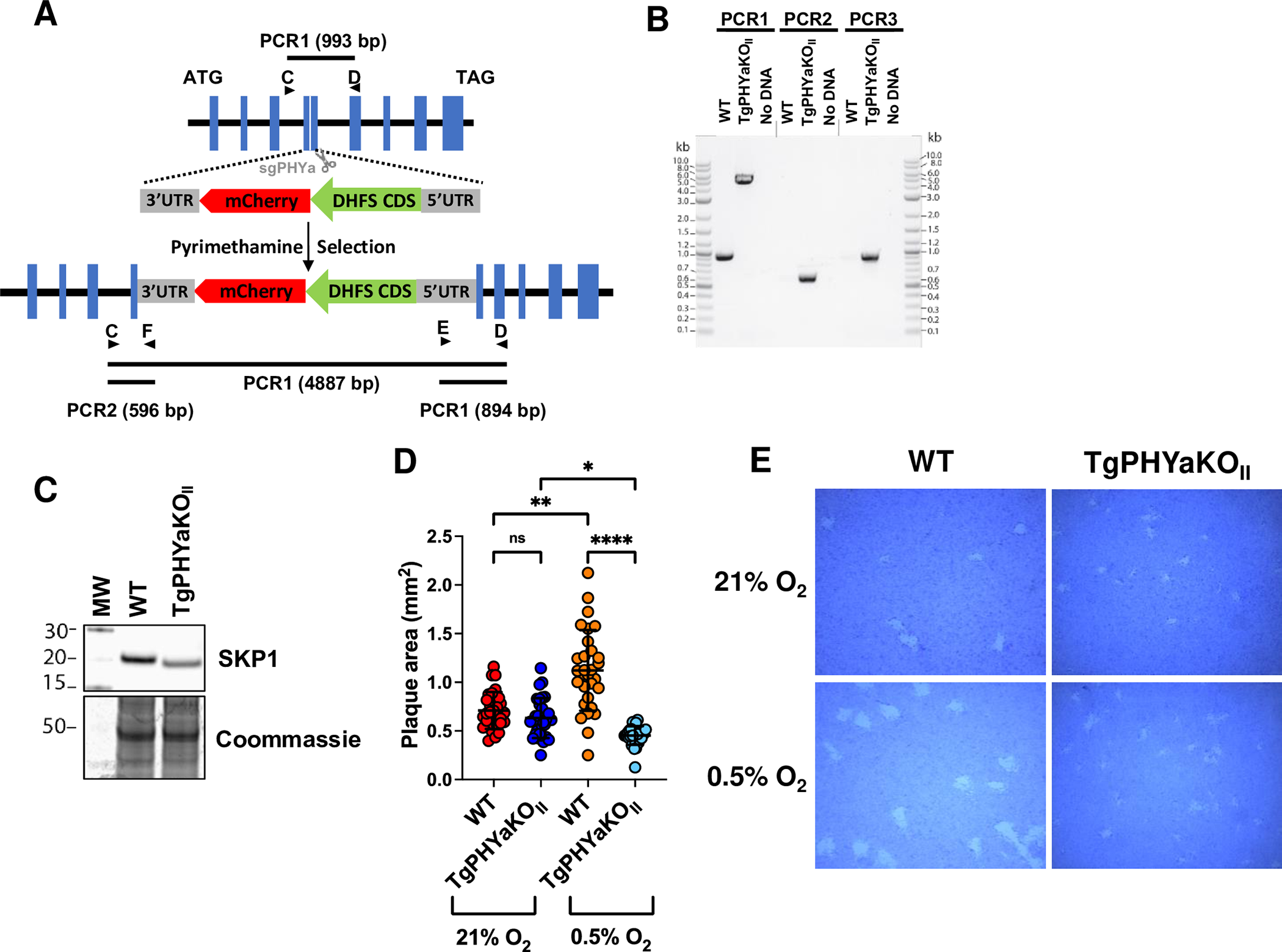
Fig 1. TgPHYa knockout in type II strain parasites.


As Toxoplasma gondii disseminates through its host, the parasite must sense and adapt to its environment and scavenge nutrients. Oxygen (O2) is one such environmental factor and cytoplasmic prolyl 4-hydroxylases (PHDs) are evolutionarily conserved O2 cellular sensing proteins that regulate responses to changes in O2 availability. Toxoplasma expresses 2 PHDs. One of them, TgPHYa hydroxylates SKP1, a subunit of the SCF-E3 ubiquitin ligase complex. In vitro, TgPHYa is important for growth at low O2 levels. However, studies have yet to examine the role that TgPHYa or any other pathogen-encoded PHD plays in virulence and disease. Using a type II ME49 Toxoplasma TgPHYa knockout, we report that TgPHYa is important for Toxoplasma virulence and brain cyst formation in mice. We further find that while TgPHYa mutant parasites can establish an infection in the gut, they are unable to efficiently disseminate to peripheral tissues because the mutant parasites are unable to survive within recruited immune cells. Since this phenotype was abrogated in IFNγ knockout mice, we studied how TgPHYa mediates survival in IFNγ-treated cells. We find that TgPHYa is not required for release of parasite-encoded effectors into host cells that neutralize anti-parasitic processes induced by IFNγ. In contrast, we find that TgPHYa is required for the parasite to scavenge tryptophan, which is an amino acid whose levels are decreased after IFNγ up-regulates the tryptophan-catabolizing enzyme, indoleamine dioxygenase (IDO). We further find, relative to wild-type mice, that IDO knockout mice display increased morbidity when infected with TgPHYa knockout parasites. Together, these data identify the first parasite mechanism for evading IFNγ-induced nutritional immunity and highlight a novel role that oxygen-sensing proteins play in pathogen growth and virulence.

Charlotte Cordonnier, Msano Mandalasi, Jason Gigley, Elizabeth A Wohlfert, Christopher M West, Ira J Blader. PLoS Biol. 2024 Jun 10;22(6):e3002690. doi: 10.1371/journal.pbio.3002690.

Massive invasion of organellar DNA drives nuclear genome evolution in Toxoplasma

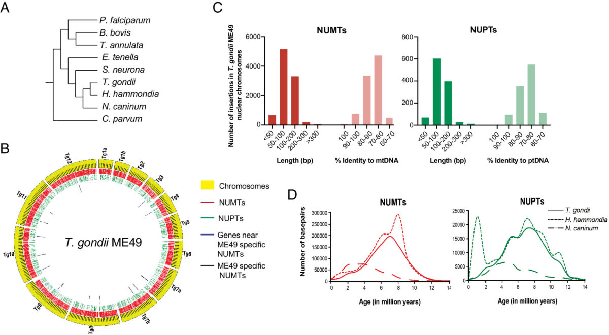
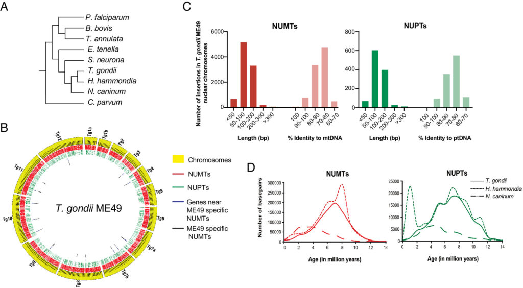
Figure 1 Characteristics of NUMTs and NUPTs in T. gondii ME49
Fig. 1 Characteristics of NUMTs and NUPTs in T. gondii ME49


Toxoplasma gondii is a zoonotic protist pathogen that infects up to one third of the human population. This apicomplexan parasite contains three genome sequences: nuclear (65 Mb); plastid organellar, ptDNA (35 kb); and mitochondrial organellar, mtDNA (5.9 kb of non-repetitive sequence). We find that the nuclear genome contains a significant amount of NUMTs (nuclear integrants of mitochondrial DNA) and NUPTs (nuclear integrants of plastid DNA) that are continuously acquired and represent a significant source of intraspecific genetic variation. NUOT (nuclear DNA of organellar origin) accretion has generated 1.6% of the extant T. gondii ME49 nuclear genome-the highest fraction ever reported in any organism. NUOTs are primarily found in organisms that retain the non-homologous end-joining repair pathway. Significant movement of organellar DNA was experimentally captured via amplicon sequencing of a CRISPR-induced double-strand break in non-homologous end-joining repair competent, but not ku80 mutant, Toxoplasma parasites. Comparisons with Neospora caninum, a species that diverged from Toxoplasma ~28 mya, revealed that the movement and fixation of five NUMTs predates the split of the two genera. This unexpected level of NUMT conservation suggests evolutionary constraint for cellular function. Most NUMT insertions reside within (60%) or nearby genes (23% within 1.5 kb), and reporter assays indicate that some NUMTs have the ability to function as cis-regulatory elements modulating gene expression. Together, these findings portray a role for organellar sequence insertion in dynamically shaping the genomic architecture and likely contributing to adaptation and phenotypic changes in this important human pathogen.

Sivaranjani Namasivayam, Cheng Sun, Assiatu B Bah, Jenna Oberstaller, Edwin Pierre-Louis, Ronald Drew Etheridge, Cedric Feschotte, Ellen J Pritham, Jessica C Kissinger. Proc Natl Acad Sci U S A. 2023 Nov 7;120(45):e2308569120. doi: 10.1073/pnas.2308569120.

Temporal and thermal profiling of the Toxoplasma proteome implicates parasite Protein Phosphatase 1 in the regulation of Ca 2+-responsive pathways

Apicomplexan parasites cause persistent mortality and morbidity worldwide through diseases including malaria, toxoplasmosis, and cryptosporidiosis. Ca2+ signaling pathways have been repurposed in these eukaryotic pathogens to regulate parasite-specific cellular processes governing the replicative and lytic phases of the infectious cycle, as well as the transition between them. Despite the presence of conserved Ca2+-responsive proteins, little is known about how specific signaling elements interact to impact pathogenesis. We mapped the Ca2+-responsive proteome of the model apicomplexan T. gondii via time-resolved phosphoproteomics and thermal proteome profiling. The waves of phosphoregulation following PKG activation and stimulated Ca2+ release corroborate known physiological changes but identify specific proteins operating in these pathways. Thermal profiling of parasite extracts identified many expected Ca2+-responsive proteins, such as parasite Ca2+-dependent protein kinases. Our approach also identified numerous Ca2+-responsive proteins that are not predicted to bind Ca2+, yet are critical components of the parasite signaling network. We characterized protein phosphatase 1 (PP1) as a Ca2+-responsive enzyme that relocalized to the parasite apex upon Ca2+ store release. Conditional depletion of PP1 revealed that the phosphatase regulates Ca2+ uptake to promote parasite motility. PP1 may thus be partly responsible for Ca2+-regulated serine/threonine phosphatase activity in apicomplexan parasites.

Alice L Herneisen, Zhu-Hong Li, Alex W Chan, Silvia N J Moreno, Sebastian Lourido. Elife. 2022 Aug 17;11:e80336. doi: 10.7554/eLife.80336.

Toxoplasma bradyzoites exhibit physiological plasticity of calcium and energy stores controlling motility and egress

Toxoplasma gondii has evolved different developmental stages for disseminating during acute infection (i.e. tachyzoites) and for establishing chronic infection (i.e. bradyzoites). Calcium ion (Ca2+) signaling tightly regulates the lytic cycle of tachyzoites by controlling microneme secretion and motility to drive egress and cell invasion. However, the roles of Ca2+ signaling pathways in bradyzoites remain largely unexplored. Here we show that Ca2+ responses are highly restricted in bradyzoites and that they fail to egress in response to agonists. Development of dual-reporter parasites revealed dampened Ca2+ responses and minimal microneme secretion by bradyzoites induced in vitro or harvested from infected mice and tested ex vivo. Ratiometric Ca2+ imaging demonstrated lower Ca2+ basal levels, reduced magnitude, and slower Ca2+ kinetics in bradyzoites compared with tachyzoites stimulated with agonists. Diminished responses in bradyzoites were associated with down-regulation of Ca2+-ATPases involved in intracellular Ca2+ storage in the endoplasmic reticulum (ER) and acidocalcisomes. Once liberated from cysts by trypsin digestion, bradyzoites incubated in glucose plus Ca2+ rapidly restored their intracellular Ca2+ and ATP stores leading to enhanced gliding. Collectively, our findings indicate that intracellular bradyzoites exhibit dampened Ca2+ signaling and lower energy levels that restrict egress, and yet upon release they rapidly respond to changes in the environment to regain motility.

Yong Fu, Kevin M Brown, Nathaniel G Jones, Silvia Nj Moreno, L David Sibley. Elife. 2021 Dec 3;10:e73011. doi: 10.7554/eLife.73011.

A terminal α3-galactose modification regulates an E3 ubiquitin ligase subunit in Toxoplasma gondii

Skp1, a subunit of E3 Skp1/Cullin-1/F-box protein ubiquitin ligases, is modified by a prolyl hydroxylase that mediates O2-regulation of the social amoeba Dictyostelium and the parasite Toxoplasma gondii. The full effect of hydroxylation requires modification of the hydroxyproline by a pentasaccharide that, in Dictyostelium, influences Skp1 structure to favor assembly of Skp1/F-box protein subcomplexes. In Toxoplasma, the presence of a contrasting penultimate sugar assembled by a different glycosyltransferase enables testing of the conformational control model. To define the final sugar and its linkage, here we identified the glycosyltransferase that completes the glycan and found that it is closely related to glycogenin, an enzyme that may prime glycogen synthesis in yeast and animals. However, the Toxoplasma enzyme catalyzes formation of a Galα1,3Glcα- rather than the Glcα1,4Glcα- linkage formed by glycogenin. Kinetic and crystallographic experiments showed that the glycosyltransferase Gat1 is specific for Skp1 in Toxoplasma and also in another protist, the crop pathogen Pythium ultimum. The fifth sugar is important for glycan function as indicated by the slow-growth phenotype of gat1Δ parasites. Computational analyses indicated that, despite the sequence difference, the Toxoplasma glycan still assumes an ordered conformation that controls Skp1 structure and revealed the importance of non-polar packing interactions of the fifth sugar. The substitution of glycosyltransferases in Toxoplasma and Pythium by an unrelated bifunctional enzyme that assembles a distinct but structurally compatible glycan in Dictyostelium is a remarkable case of convergent evolution, that emphasizes the importance of the terminal α-galactose and establishes the phylogenetic breadth of Skp1 glycoregulation.

Msano MandalasiHyun W. KimDavid ThiekerM. Osman SheikhElisabet Gas-PascualKazi RahmanPeng ZhaoNitin G. DanielHanke van der WelH. Travis IchikawaJohn N. GlushkaLance Wells, Robert J. Woods, Zachary A. Wood, and Christopher M. West. J Biol Chem. 2020 May 15. pii: jbc.RA120.013792. doi: 10.1074/jbc.RA120.013792.

Protozoan persister-like cells and drug treatment failure

Antimicrobial treatment failure threatens our ability to control infections. In addition to antimicrobial resistance, treatment failures are increasingly understood to derive from cells that survive drug treatment without selection of genetically heritable mutations. Parasitic protozoa, such as Plasmodium species that cause malaria, Toxoplasma gondii and kinetoplastid protozoa, including Trypanosoma cruzi and Leishmaniaspp., cause millions of deaths globally. These organisms can evolve drug resistance and they also exhibit phenotypic diversity, including the formation of quiescent or dormant forms that contribute to the establishment of long-term infections that are refractory to drug treatment, which we refer to as ‘persister-like cells’. In this Review, we discuss protozoan persister-like cells that have been linked to persistent infections and discuss their impact on therapeutic outcomes following drug treatment.

Michael P. Barrett, Dennis E. Kyle, L. David Sibley, Joshua B. Radke & Rick L. Tarleton. Nat Rev Microbiol. 2019 Aug 23. doi: 10.1038/s41579-019-0238-x.

Toxoplasma F-box protein 1 is required for daughter cell scaffold function during parasite replication

By binding to the adaptor protein SKP1 and serving as substrate receptors for the Skp1, Cullin, F-box E3 ubiquitin ligase complex, F-box proteins regulate critical cellular processes including cell cycle progression and membrane trafficking. While F-box proteins are conserved throughout eukaryotes and are well studied in yeast, plants, and animals, studies in parasitic protozoa are lagging. We have identified eighteen putative F-box proteins in the Toxoplasmagenome of which four have predicted homologs in Plasmodium. Two of the conserved F-box proteins were demonstrated to be important for Toxoplasma fitness and here we focus on an F-box protein, named TgFBXO1, because it is the most highly expressed by replicative tachyzoites and was also identified in an interactome screen as a Toxoplasma SKP1 binding protein. TgFBXO1 interacts with Toxoplasma SKP1 confirming it as a bona fide F-box protein. In interphase parasites, TgFBXO1 is a component of the Inner Membrane Complex (IMC), which is an organelle that underlies the plasma membrane. Early during replication, TgFBXO1 localizes to the developing daughter cell scaffold, which is the site where the daughter cell IMC and microtubules form and extend from. TgFBXO1 localization to the daughter cell scaffold required centrosome duplication but before kinetochore separation was completed. Daughter cell scaffold localization required TgFBXO1 N-myristoylation and was dependent on the small molecular weight GTPase, TgRab11b. Finally, we demonstrate that TgFBXO1 is required for parasite growth due to its function as a daughter cell scaffold effector. TgFBXO1 is the first F-box protein to be studied in apicomplexan parasites and represents the first protein demonstrated to be important for daughter cell scaffold function.

Carlos Gustavo Baptista, Agnieszka Lis, Bowen Deng, Elisabet Gas-Pascual, Ashley Dittmar, Wade Sigurdson, Christopher M. West, Ira J. Blader. PLoS Pathog. 2019 Jul 26;15(7):e1007946. doi: 10.1371/journal.ppat.1007946.

The Toxoplasma Vacuolar H+-ATPase Regulates Intracellular pH and Impacts the Maturation of Essential Secretory Proteins

Vacuolar-proton ATPases (V-ATPases) are conserved complexes that couple the hydrolysis of ATP to the pumping of protons across membranes. V-ATPases are known to play diverse roles in cellular physiology. We studied the Toxoplasma gondiiV-ATPase complex and discovered a dual role of the pump in protecting parasites against ionic stress and in the maturation of secretory proteins in endosomal-like compartments. Toxoplasma V-ATPase subunits localize to the plasma membrane and to acidic vesicles, and characterization of conditional mutants of the a1 subunit highlighted the functionality of the complex at both locations. Microneme and rhoptry proteins are required for invasion and modulation of host cells, and they traffic via endosome-like compartments in which proteolytic maturation occurs. We show that the V-ATPase supports the maturation of rhoptry and microneme proteins, and their maturases, during their traffic to their corresponding organelles. This work underscores a role for V-ATPases in regulating virulence pathways.

Andrew J.Stasic, Nathan M.Chasen, Eric J.Dykes, Stephen A.Vella, Beejan Asady, Vincent J. Starai, Silvia N.J. Moreno. 2019. Cell Rep.;27(7):2132-2146.e7. doi: 10.1016/j.celrep.2019.04.038.