Generic selectors
Exact matches only
Search in title
Search in content
Post Type Selectors

Tag: Tania Rozario

A Krüppel-like factor is required for development and regeneration of germline and yolk cells from somatic stem cells in planarians

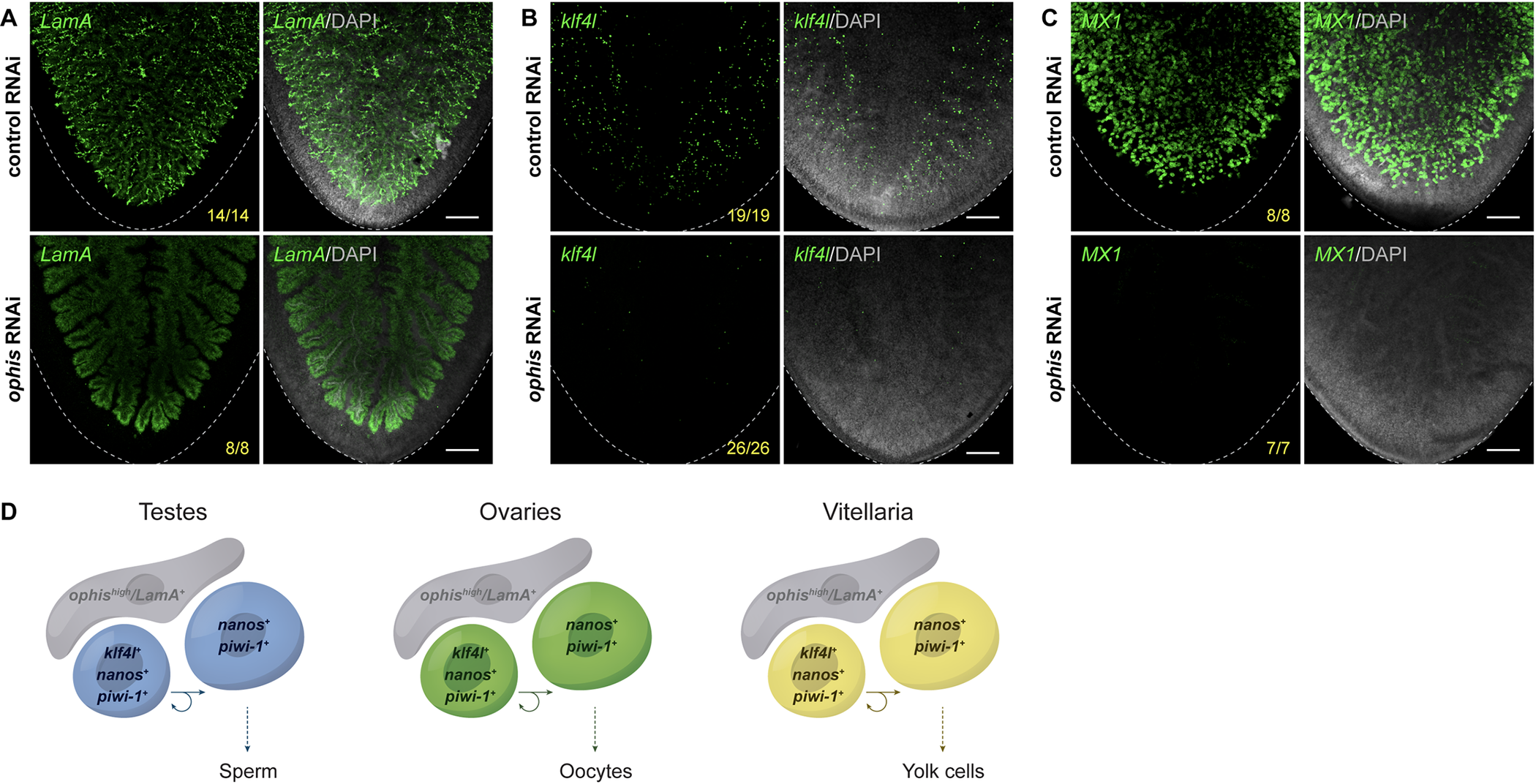
Fig 8. Germ cell niche factor ophis is required to sustain yolk cell production/vitellogenesis.
(A–C) Maximum intensity projections of confocal sections showing FISH of LamA (A), klf4l (B), and MX1 (C) (green) in the ventral posterior region of sexually mature control versus ophis RNAi animals. Dashed line denotes planarian boundary. N = 3 to 5 experiments, n = 7 to 26 planarians. (A) ophis RNAi results in a dramatic loss of the LamA+ cells throughout the vitellaria. Note that LamA expression is only visible in the branched gut in ophis RNAi planarians. (B, C) ophis RNAi results in a reduction of klf4l+ yolk cell progenitors and MX1+ differentiated yolk cells. (A–C) Nuclei are counterstained with DAPI (gray). Scale bars, 200 μm. (D) Model depicting similarities shared between gonads (where gametogenesis occurs) and vitellaria (where yolk cell production occurs). klf4l+/nanos+/piwi-1+ presumptive GSCs in testes and ovaries divide and give rise to klf4l–/nanos+/piwi-1+ progeny. These germ cells are supported by ophis+ somatic gonadal niche cells. Vitellaria are comprised of klf4l+/nanos+/piwi-1+ “germ cell–like” yolk progenitors that are mitotically competent, sustain yolk cell production, and are supported by ophishigh support cells. FISH, fluorescent RNA in situ hybridization; klf4l, klf4-like; RNAi, RNA interference.

Abstract

Sexually reproducing animals segregate their germline from their soma. In addition to gamete-producing gonads, planarian and parasitic flatworm reproduction relies on yolk cell-generating accessory reproductive organs (vitellaria) supporting development of yolkless oocytes. Despite the importance of vitellaria for flatworm reproduction (and parasite transmission), little is known about this unique evolutionary innovation. Here, we examine reproductive system development in the planarian Schmidtea mediterranea, in which pluripotent stem cells generate both somatic and germ cell lineages. We show that a homolog of the pluripotency factor Klf4 is expressed in primordial germ cells (PGCs), presumptive germline stem cells (GSCs), and yolk cell progenitors. Knockdown of this klf4-like (klf4l) gene results in animals that fail to specify or maintain germ cells; surprisingly, they also fail to maintain yolk cells. We find that yolk cells display germ cell-like attributes and that vitellaria are structurally analogous to gonads. In addition to identifying a new proliferative cell population in planarians (yolk cell progenitors) and defining its niche, our work provides evidence supporting the hypothesis that flatworm germ cells and yolk cells share a common evolutionary origin.

Melanie Issigonis, Akshada B Redkar, Tania Rozario, Umair W Khan, Rosa Mejia-Sanchez, Sylvain W Lapan, Peter W Reddien, Phillip A Newmark. PLoS Biol. 2022 Jul 15;20(7):e3001472. doi: 10.1371/journal.pbio.3001472.

The good, the bad, and the ugly: From planarians to parasites

Platyhelminthes can perhaps rightly be described as a phylum of the good, the bad, and the ugly: remarkable free-living worms that colonize land, river, and sea, which are often rife with color and can display extraordinary regenerative ability; parasitic worms like schistosomes that cause devastating disease and suffering; and monstrous tapeworms that are the stuff of nightmares. In this chapter, we will explore how our research expanded beyond free-living planarians to their gruesome parasitic cousins. We start with Schistosoma mansoni, which is not a new model; however, approaching these parasites from a developmental perspective required a reinvention that may hold generalizable lessons to basic biologists interested in pivoting to disease models. We then turn to our (re)establishment of the rat tapeworm Hymenolepis diminuta, a once-favorite model that had been largely forgotten by the molecular biology revolution. Here we tell our stories in three, first-person narratives in order to convey personal views of our experiences. Welcome to the dark side.

Tania Rozario, James J Collins 3rd, Phillip A Newmark. Curr Top Dev Biol. 2022;147:345-373. doi: 10.1016/bs.ctdb.2021.12.015.

UGA’s Rozario receives NIH Director’s New Innovator Award

By Alan Flurry

(Photo courtesy of Tania Rozario)

University of Georgia faculty member Tania Rozario has received a $2 million grant from the National Institutes of Health Director’s New Innovator Award Program, which supports early-career investigators of exceptional creativity who propose high-risk, high-reward research projects.

Rozario is an assistant professor with a joint appointment in the Franklin College of Arts and Sciences Department of Genetics and the Center for Tropical and Emerging Global Diseases.

Among the study of tropical diseases worldwide—and particularly among the parasites that cause disease—worms are a largely neglected disease agent, despite being a source of widespread problems that affect both health and economic output. Even within the study of worms, parasitic flatworms like tapeworms represent an understudied group. However, free-living flatworms like planarians are the focus of significant research because of the organism’s dynamic regenerative capacity, which presents intriguing parallels to their parasitic cousins.

Planarian flatworms cut in two will make two new worms, and cut into 10 pieces will result in 10 worms. They are the Ferrari of regenerators, according to Rozario.

“As part of its normal life cycle, a tapeworm sheds large parts of its body and then regrows this lost tissue,” Rozario said. “It has this natural regenerative-like ability, which is very promising from a basic biology standpoint, to understand how stem cells and regeneration functions in these worms.”

Taking advantage of both extensive past research and the much more sophisticated tools of today, Rozario envisions a melding of developmental biology with parasitology as a new approach to understand the parasite. She is using the rat tapeworm, Hymenolepis diminuta, to re-establish a model organism that had been a favorite model among parasitologists in the early-mid 20th century but was left behind by the molecular biology revolution.

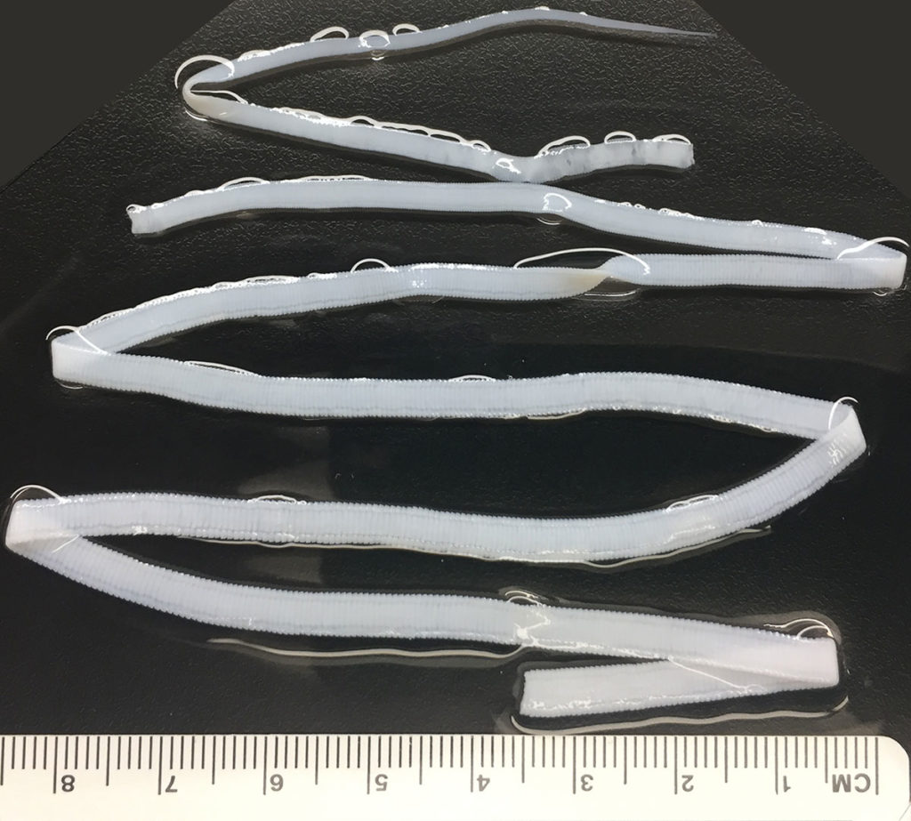
Flatworms have incredible capacity for regeneration, according to Tania Rozario, who studies them as agents of parasitic disease. For example, this rat tapeworm is capable of growing thousands of segments and can regenerate segments following amputation.


“One of the major drivers is trying to understand the diversity of the stem cell milieu in these tapeworms,” Rozario said. “We’ll try to parse out the interaction between the diversity of stem cells present and the local signals that then allow the worms to regenerate and make thousands of segments. These interactions are likely crucial for development of both female and male reproductive structures, which exist in each segment of the animal.”

“Dr. Rozario brings a new and exciting area of research to UGA, and her enthusiasm for her research is phenomenal,” said Nancy Manley, Distinguished Research Professor and department head for genetics. “Her success in getting this prestigious award speaks to her talent and the quality of her science. We are enthusiastic to have her as our newest colleague.”

“I am excited that Dr. Rozario has joined us at UGA,” said Dennis Kyle, GRA Eminent Scholar in antiparasitic drug discovery and director of the Center for Tropical and Emerging Global Diseases. “Her pioneering work is ushering in a new era whereby tapeworms can serve as model organisms. The prestigious NIH Director’s Pioneer Award is evidence of the creative approach she is undertaking to better understand these interesting parasites.”

“It’s important that we study the monsters in our midst so that we can learn from organisms in our environment that have these really out-there, unique physiological capabilities,” Rozario said. “We can learn about how they have evolved strategies to thrive in their specific niche, but they can also teach us something more fundamental about biology that could be broadly applicable.”

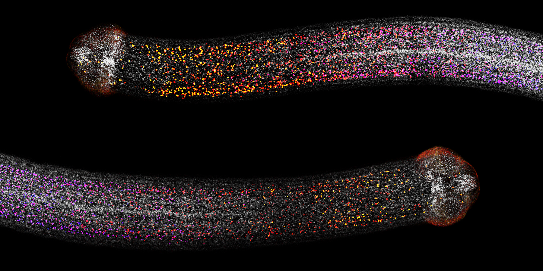
microscopy of tapeworms
In these Hymenolepis diminuta tapeworm necks, dividing cells—including stem cells—are depicted through color-marking in the image. (Image courtesy of Tania Rozario)