Protocol for detecting peptide hormones in mosquito tissues
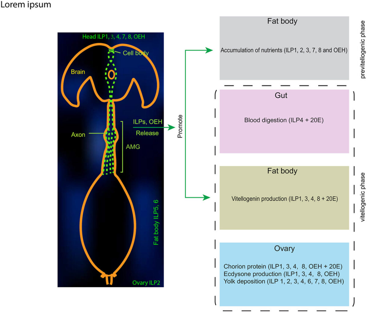
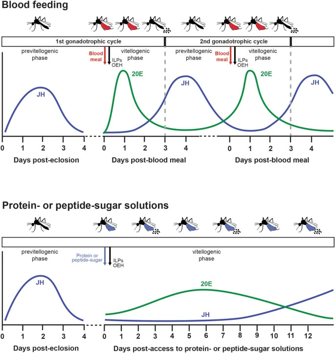
Peptide hormones in insects are primarily expressed in specialized brain, ventral nerve chord, and midgut cells. When released, peptide hormones play crucial roles in regulating physiology, reproduction, and behavior. Here, we present a protocol for detecting peptide hormones in mosquito tissues such as the brain, midgut, and hemolymph. We describe steps for tissue preparation, immunocytochemistry, …