Generic selectors
Exact matches only
Search in title
Search in content
Post Type Selectors

Tag: Diego Huet

The Toxoplasma gondii homolog of ATPase inhibitory factor 1 is critical for mitochondrial cristae maintenance and stress response

The production of energy in the form of ATP by the mitochondrial ATP synthase must be tightly controlled. One well-conserved form of regulation is mediated via ATPase inhibitory factor 1 (IF1), which governs ATP synthase activity and gene expression patterns through a cytoprotective process known as mitohormesis. In apicomplexans, the processes regulating ATP synthase activity are not fully elucidated. Using the model apicomplexan Toxoplasma gondii, we found that knockout and overexpression of TgIF1, the structural homolog of IF1, significantly affected gene expression. Additionally, TgIF1 overexpression resulted in the formation of a stable TgIF1 oligomer and increased the presence of higher order ATP synthase oligomers. We also show that parasites lacking TgIF1 exhibit reduced mitochondrial cristae density, and that while TgIF1 levels do not affect growth in conventional culture conditions, they are crucial for parasite survival under hypoxia. Interestingly, TgIF1 overexpression enhances recovery from oxidative stress, suggesting a mitohormetic function. In summary, while TgIF1 does not appear to play a role in ATP synthase regulation under conventional growth conditions, our work uncovers its potential role in adapting to the stressors faced by T. gondii and other apicomplexans throughout their intricate life cycles.

Madelaine M Usey, Anthony A Ruberto, Kaelynn V Parker, Diego Huet. Mol Biol Cell. 2024 Nov 27:mbcE24080344. doi: 10.1091/mbc.E24-08-0344.

Trainee Spotlight: Kaelynn Parker

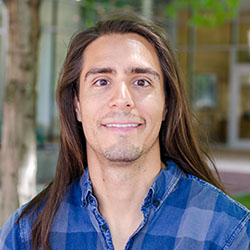
Toxoplasma background image, photo of Kaelynn Parker

I’m Kaelynn Parker and I’m from Virginia where I received my BS in biology from the University of Mary Washington. I’m a cellular biology Ph.D. student in Deigo Huet‘s laboratory.

What made you want to study science?

I took a genetic course at Germanna Community College as an elective while pursuing an associate’s degree part-time and working as an assistant barn manager. We did an experiment where we transformed E. coli with GFP and I thought it was the coolest thing I had ever done. It was a turning point where I decided I wanted to be in science.

Why did you choose UGA?

I chose UGA because of my undergraduate research advisor, Dr. Swati Agrawal, a CTEGD alum. I fell in love with parasitology (something I never imagined would happen) working with her, which inspired me to continue in the field. She also organized a seminar series featuring graduate students from CTEGD labs. After hearing from the graduate students at CTEGD, UGA was the only place I wanted to go for graduate school.

What is your project and why did you choose this research focus?

My project focuses on understanding interorganellar communication in Toxoplasma gondii through discovery and characterization of membrane contact sites between the ER, mitochondrion, and apicoplast. I am also investigating mitochondrial dynamics and stress response in T. gondii. I came to UGA with the desire to work on T. gondii because my original undergraduate project was supposed to be characterizing proteins important for egress in T. gondii. However, the COVID-19 pandemic put a halt on that plan and I wanted to return to Toxoplasma for graduate school.

What are your career goals?

I would like to remain in academic parasitology.

What do you hope to do for your capstone experience? Is there a collaborator/field site you would like to visit?

For my capstone experience, my plan is to utilize the opportunity to go to another lab to learn techniques to apply to membrane contact site research.

What is your favorite thing about UGA and/or Athens?

I love to go bird watching at the botanical gardens and local parks.

Any advice for a student interested in this field?

Talk to people, take every opportunity to present your work and build connections.


Support trainees like Kaelynn by giving today to the Center for Tropical & Emerging Global Diseases.

Seeing the unseen: illuminating Toxoplasma gondii’s metabolic manipulation

Intracellular infection by a pathogen induces significant rewiring of host cell signaling and biological processes. Understanding how an intracellular pathogen such as Toxoplasma gondii modulates host cell metabolism with single-cell resolution has been challenged by the variability of infection within cultures and difficulties in separating host and parasite metabolic processes. A new study from Gallego-Lopez and colleagues (G. M. Gallego-López, E. C. Guzman, D. E. Desa, L. J. Knoll, M. C. Skala, mBio e00727-24, 2024, https://doi.org/10.1128/mbio.00727-24) applies a quantitative imaging approach to evaluate the host cell metabolism during intracellular infection with Toxoplasma. This study provides important insights into host metabolic responses to Toxoplasma infection and offers a valuable tool to dissect the mechanisms underlying parasite infection and pathophysiology.

Diego Huet, Victoria Jeffers. mBio. 2024 Jul 12:e0121124. doi: 10.1128/mbio.01211-24.

Diego Huet zeroes in on parasite that affects thousands each year

by Donna Huber

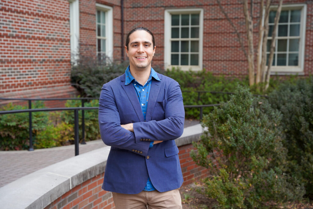
Diego Huet
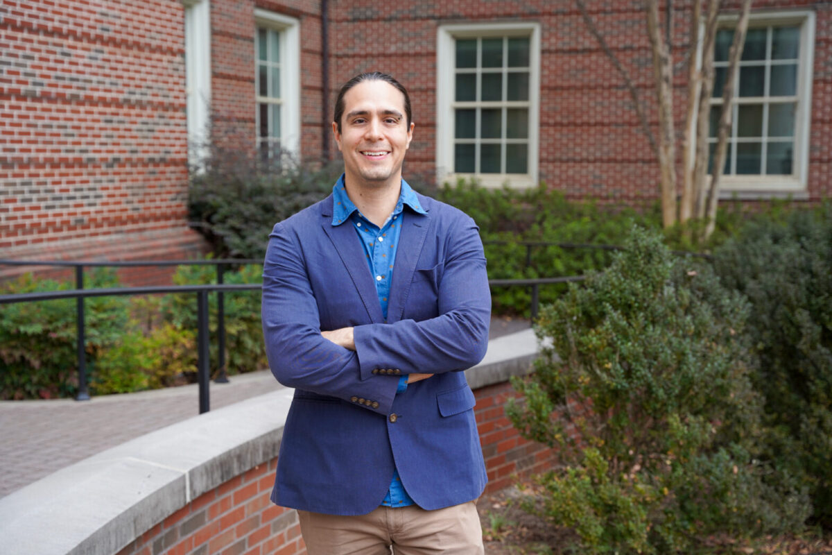
Diego Huet, assistant professor in the College of Pharmacy and the Center for Tropical & Emerging Global Diseases, studies parasites that cause disease in both humans and animals. His lab has ramped up a project to better understand the biology of Toxoplasma gondii , an organism carried by cats that is related to the parasite that causes malaria. (Photo by Lauren Corcino)


From an early age, Diego Huet has been interested in the unusual and fascinating found in the natural world.

His early encounters with animals, plants and insects nurtured his curiosity about nature. Their striking colors and sometimes strange shapes drew his interest, and even today he continues to capture them through macro photography. It was this fascination that led him to the parasite he studies today.

“I was always drawn to ‘unconventional’ or ‘weird’ science,” said Huet, an assistant professor in the College of Pharmacy’s Department of Pharmaceutical and Biomedical Sciences and member of the Center for Tropical and Emerging Global Diseases. “But I also wanted to be hands on, which is what led me to molecular and cellular biology.”

As a doctoral student, Huet began studying Trypanosoma brucei, a parasite commonly transmitted by the tsetse fly. Wanting to study a different parasite as a postdoctoral researcher, he was torn between studying Plasmodium, which is the causative agent of malaria, and Toxoplasma gondii, a related parasite that is carried by cats. Both parasites, which belong to a group of organisms called apicomplexans, cause diseases in humans and animals, and there remain large knowledge gaps in our basic understanding of them. Ultimately, he chose the latter.

Plasmodium is difficult to manipulate,” Huet said. “Toxoplasma is related to Plasmodium, but is easier to work with because it isn’t as complex, and what we learn about Toxo could also increase our knowledge of Plasmodium.”

Just as yeast and fruit flies are used as model organisms to study human biology, Toxoplasma can be used as a model for shared features of apicomplexan biology.

Besides aiding in the understanding of other parasites of human and veterinary concern, including parasites that cause malaria in tropical and subtropical regions of the world, Toxoplasma gondii also causes human and animal disease. More than 40 million people in the U.S. are estimated to carry T. gondii. Although most never show symptoms, it poses a major health threat to immunocompromised individuals and pregnant women as it can lead to miscarriage and birth defects. Toxoplasmosis, the disease caused by Toxoplasma, is considered a leading cause of death among foodborne illnesses though it can also be transmitted through contact with cat feces.

The Centers for Disease Control and Prevention has it listed as a neglected parasitic infection in the United States and a target for public health action.

Huet joined the faculty at the University of Georgia in 2019 and has developed a robust research program to expand knowledge of the basic biology of Toxoplasma.
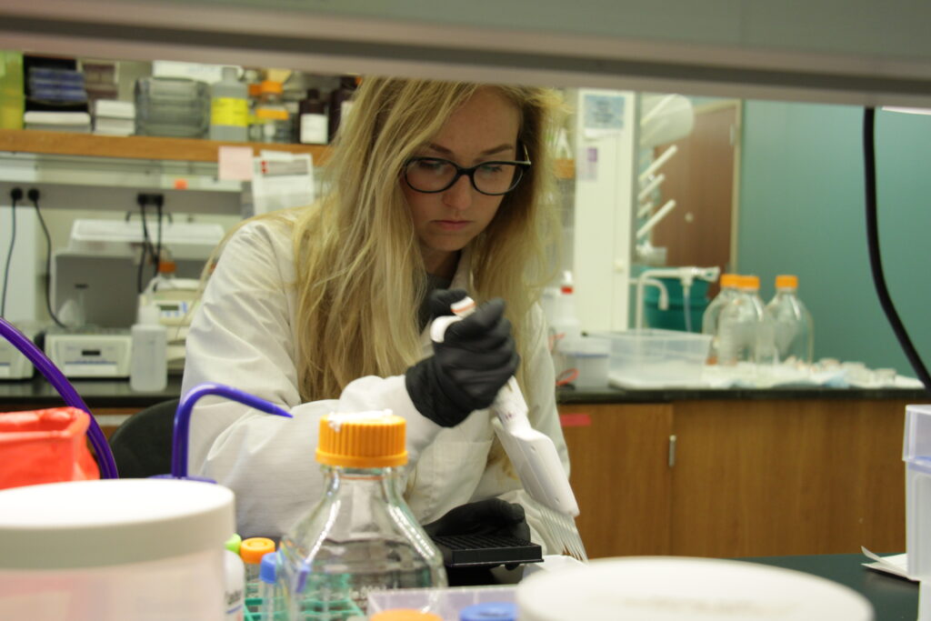
Madelaine Usey
Madelaine Usey is a cellular biology graduate student in the Huet Laboratory.

In a recently published study in “mBio”, cellular biology doctoral candidate and Huet Lab member Madelaine Usey looked at proteins critical for mitochondrial function in T. gondii. The mitochondrion is considered the “powerhouse of the cell,” but it is an enzyme called ATP synthase that generates the cellular energy.

“Our findings are really exciting for drug discovery,” Usey said. “Many of the proteins that make up the ATP synthase are different in Toxoplasma compared to other organisms. In this study, we were able to figure out what two of those novel subunits are doing—they act as scaffolding for this enormous ATP synthase complex.”

These proteins are unique to Toxoplasma and could be used in drug discovery as targets since they are important for mitochondrial functioning.

Another project in Huet’s laboratory, which recently received funding through a grant from the National Institute of General Medical Sciences, investigates how organelles within the parasite communicate.

“Traditionally, we thought organelles send and receive calcium and other metabolites in much the same way we receive a package through the mail,” Huet said. “Cells form vesicles to transport materials to specific locations within the cell. The vesicles are labeled with proteins that act like a postal address, telling the vesicle where to go.”

However, cells can also exchange material through another process.

“When the organelles’ membranes get close together, they form what is called a membrane contact site,” Huet said. “In this case it is more like one organelle hand delivers the package to another.”

A membrane contact site is a specialized protein structure that organelles use for intracellular communication. However, it is not a well understood structure in apicomplexans. In addition, these parasites have additional organelles not found in traditional models like humans and yeast, so Huet is trying to understand how the organellar communication is happening in apicomplexans using Toxoplasma as a model.

Identifying such proteins and their functions could lead to better drug targets and better drug treatments, which all the neglected parasitic diseases need.

“Toxo’s genome isn’t well annotated,” Huet said. “Finding membrane contact site proteins is an arduous task—it’s a goal of my lab to identify some of them and their involvement in Toxoplasma membrane contact sites.”


This article was first published at https://research.uga.edu/news/diego-huet-zeroes-in-on-parasite-that-affects-thousands-each-year/

ATP synthase-associated coiled-coil-helix-coiled-coil-helix (CHCH) domain-containing proteins are critical for mitochondrial function in Toxoplasma gondii

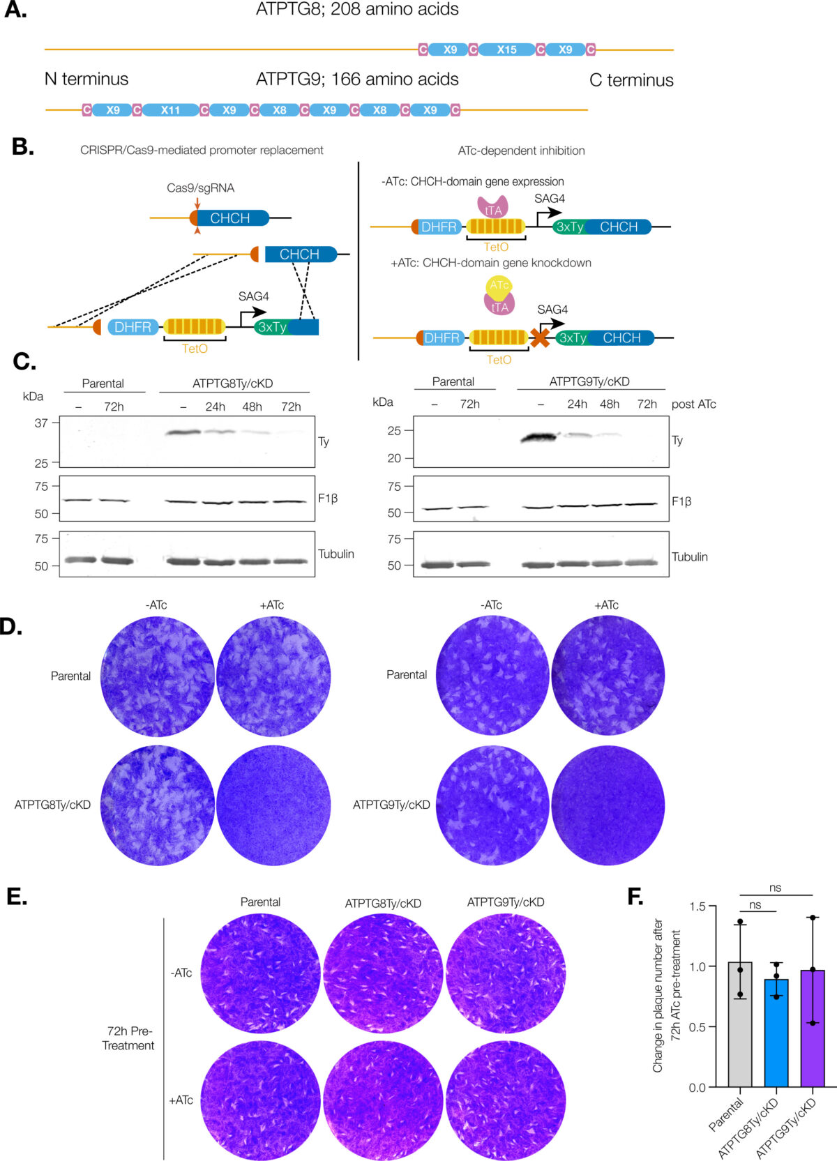
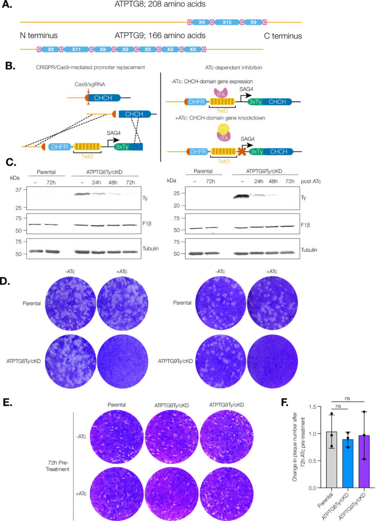
CHCH domain proteins associated with the T. gondii ATP synthase are essential for the lytic cycle. (A) Schematic representation of the CHCH domain size and location in ATPTG8 and ATPTG9. C represents cysteine residues and X represents any other amino acid residue.

Coiled-coil-helix-coiled-coil-helix (CHCH) domains consist of two pairs of cysteine residues that are oxidized to form disulfide bonds upon mitochondrial import. Proteins containing these domains play important roles in mitochondrial ultrastructure and in the biogenesis, function, and stability of electron transport chain complexes. Interestingly, recent investigations of the Toxoplasma gondii ATP synthase identified subunits containing CHCH domains. As CHCH domain proteins have never been found in any other ATP synthase, their role in T. gondii was unclear. Using conditional gene knockdown systems, we investigated two T. gondii ATP synthase subunits containing CHCH domains: ATPTG8 and ATPTG9. We show that these two subunits are essential for the lytic cycle as well as stability and function of the ATP synthase. Further, we illustrated that their knockdown disrupts multiple aspects of mitochondrial morphology, including ultrastructure and cristae density. Mutation of key cysteine residues in the CHCH domains also caused mis-localization of the proteins. Our work suggests that these proteins likely provide structural support to the exceptionally large T. gondii ATP synthase complex and that perturbations to the structural integrity of this complex result in deleterious downstream effects on the parasite mitochondrion. These investigations add to a growing body of work focused on the divergent aspects of the apicomplexan ATP synthase, which could ultimately uncover novel drug targets. IMPORTANCE Members of the coiled-coil-helix-coiled-coil-helix (CHCH) domain protein family are transported into the mitochondrial intermembrane space, where they play important roles in the biogenesis and function of the organelle. Unexpectedly, the ATP synthase of the apicomplexan Toxoplasma gondii harbors CHCH domain-containing subunits of unknown function. As no other ATP synthase studied to date contains this class of proteins, characterizing their function will be of broad interest to the fields of molecular parasitology and mitochondrial evolution. Here, we demonstrate that that two T. gondii ATP synthase subunits containing CHCH domains are required for parasite survival and for stability and function of the ATP synthase. We also show that knockdown disrupts multiple aspects of the mitochondrial morphology of T. gondii and that mutation of key residues in the CHCH domains caused mis-localization of the proteins. This work provides insight into the unique features of the apicomplexan ATP synthase, which could help to develop therapeutic interventions against this parasite and other apicomplexans, such as the malaria-causing parasite Plasmodium falciparum.

Madelaine M Usey, Diego Huet. mBio. 2023 Oct 5:e0176923. doi: 10.1128/mbio.01769-23.

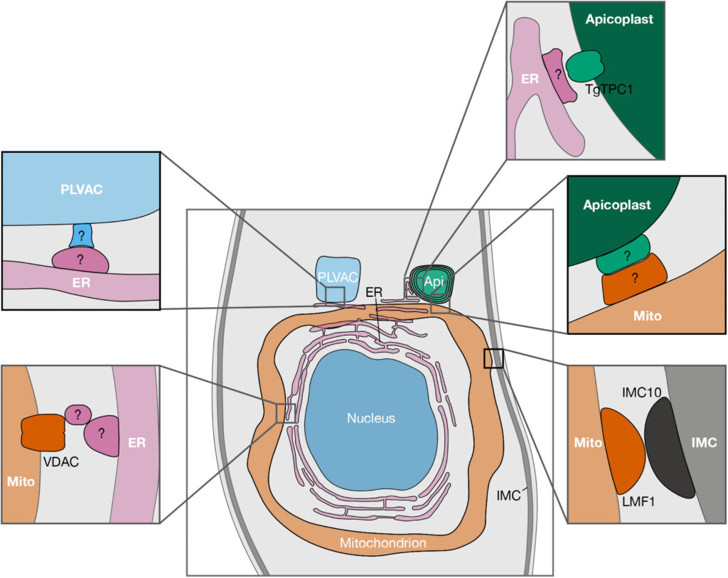
Interorganellar Communication Through Membrane Contact Sites in Toxoplasma gondii

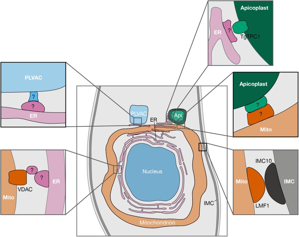
Figure 1. Reported and potential MCSs between organelles of Toxoplasma gondii. Schematic representation showing proteins recently reported to be involved in MCSs, along with putative MCS candidates (indicated with “?”). For clarity purposes, only the central part of the parasite is shown. Abbreviations: AP, apicoplast; ER, endoplasmic reticulum; PLVAC, plant-like vacuolar compartment; IMC, inner membrane complex; TgTPC, T. gondii two pore channel; VDAC, voltage-dependent anion channel; LMF1, lasso maintenance factor 1.; MCS, membrane contact site.


Apicomplexan parasites are a group of protists that cause disease in humans and include pathogens like Plasmodium spp., the causative agent of malaria, and Toxoplasma gondii, the etiological agent of toxoplasmosis and one of the most ubiquitous human parasites in the world. Membrane contact sites (MCSs) are widespread structures within eukaryotic cells but their characterization in apicomplexan parasites is only in its very beginnings. Basic biological features of the T. gondii parasitic cycle support numerous organellar interactions, including the transfer of Ca2+ and metabolites between different compartments. In T. gondii, Ca2+ signals precede a series of interrelated molecular processes occurring in a coordinated manner that culminate in the stimulation of key steps of the parasite life cycle. Calcium transfer from the endoplasmic reticulum to other organelles via MCSs would explain the precision, speed, and efficiency that is needed during the lytic cycle of T. gondii. In this short review, we discuss the implications of these structures in cellular signaling, with an emphasis on their potential role in Ca2+ signaling.

Diego Huet, Silvia N J Moreno. Contact (Thousand Oaks). 2023 Aug 6;6:25152564231189064. doi: 10.1177/25152564231189064. eCollection 2023 Jan-Dec.

The mystery of massive mitochondrial complexes: the apicomplexan respiratory chain

The mitochondrial respiratory chain is an essential pathway in most studied eukaryotes due to its roles in respiration and other pathways that depend on mitochondrial membrane potential. Apicomplexans are unicellular eukaryotes whose members have an impact on global health. The respiratory chain is a drug target for some members of this group, notably the malaria-causing Plasmodium spp. This has motivated studies of the respiratory chain in apicomplexan parasites, primarily Toxoplasma gondii and Plasmodium spp. for which experimental tools are most advanced. Studies of the respiratory complexes in these organisms revealed numerous novel features, including expansion of complex size. The divergence of apicomplexan mitochondria from commonly studied models highlights the diversity of mitochondrial form and function across eukaryotic life.

Andrew E Maclean, Jenni A Hayward, Diego Huet, Giel G van Dooren, Lilach Sheiner. Trends Parasitol. 2022 Oct 24;S1471-4922(22)00219-7. doi: 10.1016/j.pt.2022.09.008. Online ahead of print.

Parasite Powerhouse: a Review of the Toxoplasma gondii Mitochondrion

Toxoplasma gondii is a member of the apicomplexan phylum, a group of single-celled eukaryotic parasites that cause significant human morbidity and mortality around the world. T. gondii harbors two organelles of endosymbiotic origin: a non-photosynthetic plastid, known as the apicoplast, and a single mitochondrion derived from the ancient engulfment of an α-proteobacterium. Due to excitement surrounding the novelty of the apicoplast, the T. gondii mitochondrion was, to a certain extent, overlooked for about two decades. However, recent work has illustrated that the mitochondrion is an essential hub of apicomplexan-specific biology. Development of novel techniques, such as cryo-electron microscopy, complexome profiling, and next-generation sequencing have led to a renaissance in mitochondrial studies. This review will cover what is currently known about key features of the T. gondii mitochondrion, ranging from its genome to protein import machinery and biochemical pathways. Particular focus will be given to mitochondrial features that diverge significantly from the mammalian host, along with discussion of this important organelle as a drug target.

Madelaine M. Usey, Diego Huet. J Eukaryot Microbiol. 2022 Mar 21;e12906. doi: 10.1111/jeu.12906.

Trainee Spotlight: Megna Tiwari

Megna Tiwari 

Megna Tiwari is a second-year Ph.D. trainee in the laboratory of Diego Huet. She is originally from Newport Beach, California and completed her undergraduate degree in Cell, Molecular and Developmental Biology at the University of California, Riverside (UCR). While at UCR, she worked as an undergraduate researcher in the fungal genomics lab of Dr. Jason Stajich for 2 years and co-founded Women in STEM Engaging Riverside (WISER). After graduation, she worked as a blood bank lab technician at LifeStream Blood Bank where she screened for and routinely found blood samples positive for understudied pathogenic parasites. Her fascination with pathogenic parasites led her to seek a thesis-based Master of Science in Biology at California State University, Fullerton under the supervision of Dr. Veronica Jimenez. During this period, Megna worked on understanding the functional and structural relationship of mechanosensitive ion channels found in T. cruzi and cemented her passion for molecular parasitology.

Megna has been awarded a CTEGD T32 Training Fellowship. She currently serves as Vice-president of CTEGD’s Graduate Student Association and New Student Liaison for the Department of Cellular Biology’s Graduate Student Association.

Why did you choose UGA? 

My master’s research in parasitology reaffirmed my passion for research in unconventional parasitic pathogens. Therefore, I applied for doctoral programs that would allow me to remain in the field of cell and molecular parasitology and the CTEGD at UGA was the perfect place for me to obtain the best possible training as a parasitologist.

What is your research focus/project and why are you interested in the topic? 

The over-reaching research goal of the Huet lab is the investigation of the highly divergent metabolic adaptations of apicomplexans. My research interests in the lab have led me to study the role of the ATP synthase in the apicomplexan Toxoplasma gondii, the causative agent of toxoplasmosis. For my project, I am examining the role of apicomplexan-specific ATP synthase subunits and how they might contribute to the regulation of the ATP synthase function in the parasite.

What are your future professional plans?  

Following graduation from UGA, I hope to continue on for a postdoctoral research position in parasitology.

What do you hope to do for your capstone experience? 

For my capstone experience, I want to gain an outside perspective and understanding of foreign research culture that I can apply to my own research when I return to the CTEGD. 

What is your favorite thing about UGA and/or Athens? 

At the CTEGD, I love the collaborative nature. If I am trying to learn a new technique or understand new concepts, I am able to easily walk down the hall to a neighboring lab and get advice. In Athens, for entertainment, I love the endless craft beer scene and I love all the greenery and being able to hike gaps of the Appalachian trail!


Support trainees like Megna by giving today to the Center for Tropical & Emerging Global Diseases.

[button size=’large’ style=” text=’Give Now’ icon=” icon_color=’BA0C2F’ link=’https://ctegd.uga.edu/give/’ target=’_self’ color=” hover_color=” border_color=” hover_border_color=” background_color=” hover_background_color=” font_style=” font_weight=” text_align=’center’ margin=”]