Chet Joyner receives $1.1 million grant to study malaria vaccine

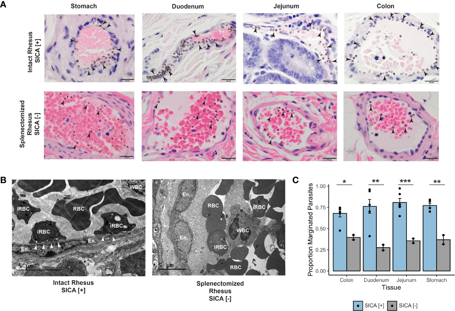
RESEARCH WILL BE IN COLLABORATION WITH YALE UNIVERSITY Chet Joyner, PhD, a faculty member in the Center for Vaccines and Immunology and the Center for Tropical and Emerging Diseases in the College of Veterinary Medicine (CVM) at the University of Georgia, is the recipient of a $1.1 million grant from Open Philanthropy to perform preclinical …