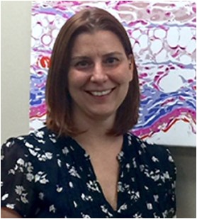
Invited Speaker Spotlight: Tiffany Weinkoff

About the Speaker
 Macrophages are the major cell type infected by Leishmania parasites and the goal of my research is to define the relationship between Leishmania parasites, macrophages and the vasculature. Throughout my graduate studies and postdoctoral positions, I have dedicated my efforts toward understanding parasite-host interactions. As a graduate student at the University of Georgia carrying out research at the Centers for Disease Control and Prevention under Dr. Patrick Lammie, I was exposed to high caliber basic research in the CTEGD while simultaneously confronted with important public health issues in field settings related to neglected tropical diseases. My predoctoral research focused on the ability of monocytes to modulate lymphatic endothelial cell function and how this interaction contributed to the pathogenesis of human filarial disease. For my first postdoctoral position, I moved to the laboratory of Dr. Fabienne Tacchini-Cottier at the University of Lausanne in Switzerland. In Switzerland I gained experience using the mouse model to study the immunologic mechanisms involved in susceptibility to Leishmania infection. For my second postdoctoral position, I went to the laboratory of Phillip Scott at the University of Pennsylvania, a world-renowned professor and institution in the field of immunoparasitology. My initial project in the Scott lab, addressed the role of M2 macrophages as safe havens during Leishmania infection. For my second project in the Scott lab, I merged the knowledge and experience gained from my predoctoral research studying myeloid cells and vessels in filariasis with the technical experience of my first postdoctoral position to address the roles of innate cells in vivo in leishmaniasis. In the past months, I have transitioned to an Assistant Professor at the University of Arkansas for Medical Sciences (UAMS) where I have joined an elite group of vibrant and enthusiastic scientists with expertise in host-pathogen interactions. At UAMS, I am expanding upon previous studies examining vascular remodeling and the role of the VEGF-A/VEGFR-2 signaling pathway in lymphangiogenesis during Leishmania infection. In the future, I will work in collaboration with Dr. Camila Oliveira in Bahia, Brazil in a new project focusing on vascular remodeling during murine and human Leishmania braziliensis infection. These studies will provide important insights into our current understanding of the role of the vasculature during human disease.
Tiffany Weinkoff’s Talk
Dr. Weinkoff will give the following talk at 2:45 pm
The Role of Myeloid Cells in Vascular Remodeling during Leishmania major Infection
Tiffany Weinkopff
Department of Microbiology and Immunology, College of Medicine, University of Arkansas for Medical Sciences, Little Rock, AR, 72205, USA
Our lab investigates the mechanistic basis for the pathogenesis of disease driven by Leishmania, a protozoal parasite that causes cutaneous lesions. Following infection, lesion severity is often increased by an exaggerated inflammatory response. As a result, the inflammatory response can maintain disease even after the parasite infection has been controlled. Given vascular remodeling contributes to the magnitude of many inflammatory conditions, it is hypothesized that the manipulation of factors promoting angiogenesis or lymphangiogenesis would alter lesion severity in leishmaniasis. Our findings demonstrate that murine Leishmania major infection leads to dramatic changes in vessel morphology, number and permeability. At the peak of infection, VEGF-A and VEGFR-2 expression are upregulated and VEGFR-2 blockade led to a reduction in lymphatic endothelial cell proliferation and simultaneously increased lesion size without altering the parasite burden. We showed VEGF-A/VEGFR-2 signaling promotes lymphangiogenesis to restrict tissue inflammation in leishmaniasis. Given VEGF-A/VEGFR-2 signaling contributes to lesion resolution, we are currently investigating the cellular and molecular mediators driving VEGF-A production. We found that macrophages are the predominant cell type expressing VEGF-A during L. major infection and that parasites can directly induce VEGF-A production by macrophages in vitro. Given Leishmania parasites activate HIF-1α and this transcription factor induces VEGF-A expression, we analyzed the expression of HIF-1α during infection. We showed that macrophages are the major cell type expressing HIF-1α during infection and that parasite-induced VEGF-A production is mediated by HIF activation. We are presently examining VEGF-A expression in mice deficient in HIF signaling specifically in myeloid cells. To date, we have shown that LysMCre ARNTf/f mice express less VEGF-A than LysMCre ARNTf/+ control mice following infection, and we are exploring how decreased myeloid VEGF-A production influences vascular remodeling and lesion resolution in these animals. Altogether, these studies suggest macrophage HIF-dependent VEGF-A production contributes to lymphatic remodeling during L. major infection.
More information about the Molecular Parasitology & Vector Biology Symposium and the schedule of presentions are available on our website. The deadline to register for the symposium is April 24.
